Gallbladder disease is one of the most common conditions I treat as a general surgeon. An estimated 20 million Americans have gallstones, and removing the gallbladder, a procedure called a cholecystectomy, is one of the most frequently performed surgeries in the country.¹ That is exactly why I am starting this series here.
When you come to my office for a consultation, before I start talking about how a procedure is done, I walk you through what the structures look like, how they work, and what is actually going wrong. I am going to do the same thing here on the blog. Each topic in this series will follow that same order: anatomy, then physiology, then pathology, then surgery. First up: the gallbladder and the biliary tree.
What Is the Biliary Tree?
The word "biliary" refers to bile, the digestive fluid your liver makes every day. The biliary tree is the branching network of tubes that carries bile from your liver to your small intestine. Think of it like the plumbing under a sink: there are main pipes, smaller branches, and a storage tank. The gallbladder is that storage tank.
The Liver: Where It All Starts
Your liver makes bile continuously, around the clock, whether you are eating or not. Bile drains out of the liver through small tubes called the right and left hepatic ducts. These two ducts come together to form a single larger tube called the common hepatic duct, which carries bile downward and out of the liver.²
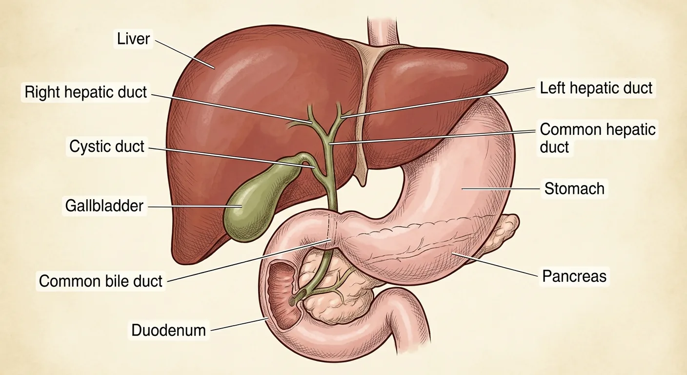
Image: normal biliary anatomy. Liver, hepatic ducts, gallbladder, cystic duct, common bile duct, pancreatic duct, and duodenum.
The Gallbladder: The Storage Tank
The gallbladder is a small, pear-shaped organ tucked right up under the liver on your right side. It connects to the common hepatic duct through its own separate tube called the cystic duct. Between meals, bile backs up through the cystic duct and gets stored inside the gallbladder. The gallbladder also concentrates the bile while it holds it, making it stronger and more effective for when you actually need it.²
The Common Bile Duct
Below the point where the cystic duct branches off, the main bile tube continues downward under a new name: the common bile duct. This carries bile from the liver and gallbladder the rest of the way down toward the small intestine.²
Calot's Triangle: The Most Important Landmark in Gallbladder Surgery
The area where the cystic duct and the common hepatic duct come together near the undersurface of the liver is known as Calot's triangle. It was first described in 1891 by the French surgeon Jean-François Calot in his doctoral thesis. Calot originally described it as a triangle formed by the cystic duct, the common hepatic duct, and the cystic artery.³
It is worth knowing that the modern surgical definition has evolved from Calot's original description. Today, surgeons define the superior border of this triangle as the undersurface of the liver rather than the cystic artery. This updated version is sometimes called the hepatocystic triangle.³ The current boundaries are the cystic duct on one side, the common hepatic duct on another, and the undersurface of the liver above.³
Regardless of how the borders are defined, this small space is one of the most critical landmarks in gallbladder surgery. The cystic artery, which supplies blood to the gallbladder, runs through this triangle. Before I can safely remove the gallbladder, I have to clearly identify and confirm every structure inside this space. Careful dissection here is essential to avoid injury to the bile ducts and nearby blood vessels.³,⁴,⁵ We will come back to this in detail when we cover the surgery later in this series.
Where Bile and Pancreatic Juice Meet
The common bile duct does not enter the small intestine on its own. Just before it gets there, it joins the pancreatic duct, which carries digestive juices from the pancreas. These two ducts come together at a shared opening called the ampulla of Vater, and both bile and pancreatic juice enter the duodenum, the first part of the small intestine, through this single point.²
This connection matters clinically. A gallstone that travels out of the gallbladder and gets stuck at or near the ampulla of Vater can block both bile flow and pancreatic drainage at the same time. That is one reason gallstone disease can sometimes cause pancreatitis. We will cover that in detail later in this series.
The Key Structures to Know
Here is your map of the biliary tree:
- Liver: makes bile continuously
- Right and left hepatic ducts: carry bile out of the liver
- Common hepatic duct: the two ducts joined together
- Gallbladder: stores and concentrates bile between meals
- Cystic duct: connects the gallbladder to the main bile duct
- Common bile duct: carries bile from the liver and gallbladder to the intestine
- Pancreatic duct: carries digestive juice from the pancreas
- Ampulla of Vater: the shared opening where bile and pancreatic juice enter the duodenum together
Understanding where each one sits, and how they connect, is the foundation for everything else in this series.
Up next: what this biliary tree actually does. How bile is made, stored, and released to help you digest fat.
References
1. Laparoscopic Cholecystectomy. StatPearls [Internet]. Treasure Island, FL: StatPearls Publishing.
2. Jones M, Hannoodee S. Anatomy, Abdomen and Pelvis, Gallbladder. StatPearls [Internet]. Treasure Island, FL: StatPearls Publishing.
3. Abdalla S, Pierre S, Ellis H. Calot's Triangle. Clinical Anatomy. 2013.
4. Suzuki M, Akaishi S, Rikiyama T, et al. Laparoscopic Cholecystectomy, Calot's Triangle, and Variations in Cystic Arterial Supply. Surgical Endoscopy. 2000.
5. Bergamaschi R, Ignjatovic D. More Than Two Structures in Calot's Triangle. A Postmortem Study. Surgical Endoscopy. 2000.