The short version
- One incision (about 10 cm) in the groin crease; it can be done under local anesthesia with sedation, a spinal block, or general anesthesia.
- The groin nerves are protected throughout the repair to lower the risk of numbness or chronic pain.
- The bulging tissue is returned to place and the weak spot is reinforced with .
- Most patients go home the same day; nothing over 10 pounds until your 2 week visit, when we reassess.
A quick summary. The full details are below, and none of it replaces an exam.
What is an inguinal hernia?
An hernia happens when tissue, often part of the intestine or surrounding fat, pushes through a weak spot in your lower abdominal wall, in the groin area. This creates a bulge, and often discomfort or pain, especially when lifting, coughing, or straining. Not every ache in that area means you have a hernia, which is why an exam matters.
Why does this happen?
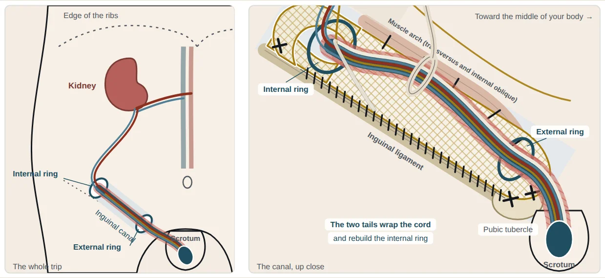
The groin area naturally has a passage called the inguinal canal. While a baby is developing, this canal is where the testicles travel down into the scrotum (in males). That process leaves a natural weak spot in the abdominal wall for everyone, which can widen over time or under strain.
Three types of hernia in this area
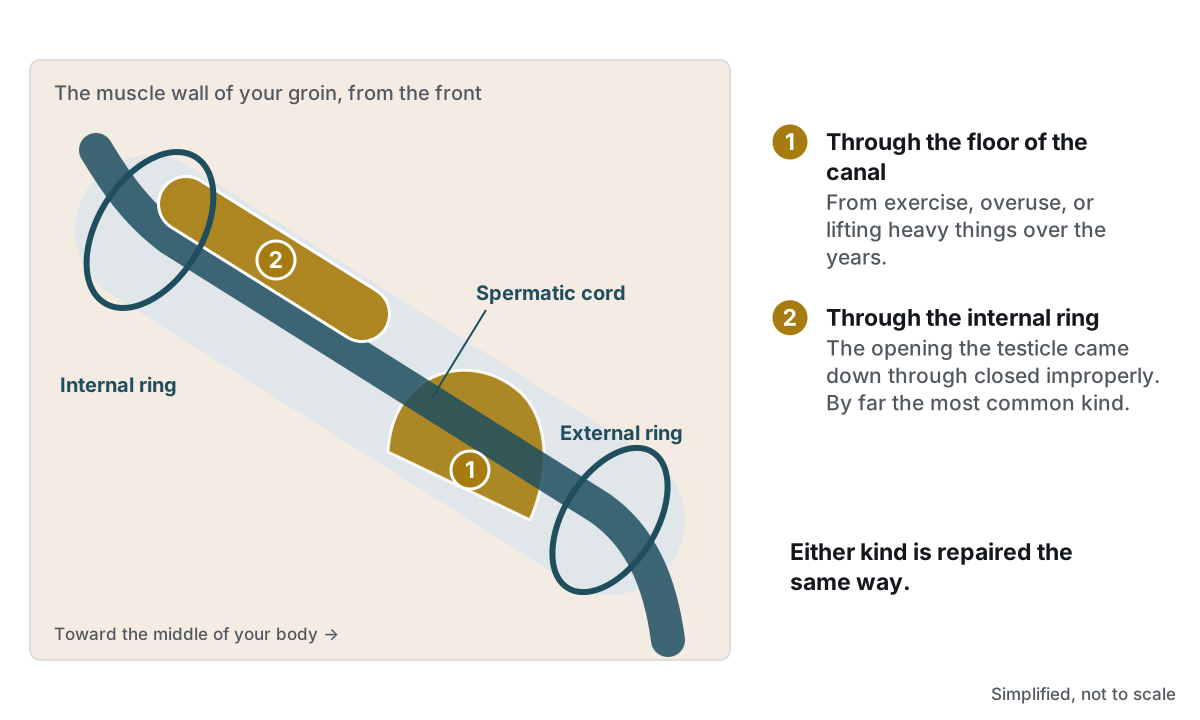
- Indirect inguinal hernia: the most common type. Often present from birth, it follows the same path the testicles took during development.
- Direct inguinal hernia: develops later in life from general wear and weakening of the abdominal wall muscles.
- : less common, and located just below the main inguinal area, near the blood vessels that travel into the thigh. This type is more common in women.
How open repair works
Open inguinal hernia repair is done through a single incision in the groin, directly over the hernia, rather than through small ports in the abdomen. Depending on your health and preference, it can be done under local anesthesia with sedation, a spinal block, or general anesthesia. We'll discuss which option fits you best.
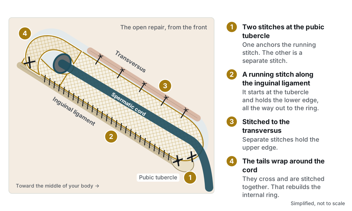
This is a repair, not a removal. The hernia itself isn't taken out. Instead, the bulging tissue is reduced back into place, and the weak spot is reinforced with mesh to keep it from coming back.
Step by step
- Getting in: a single incision, about 10cm long, is made in the groin crease directly over the hernia.
- Opening the inguinal canal: the layers of tissue are opened to expose the inguinal canal and the .
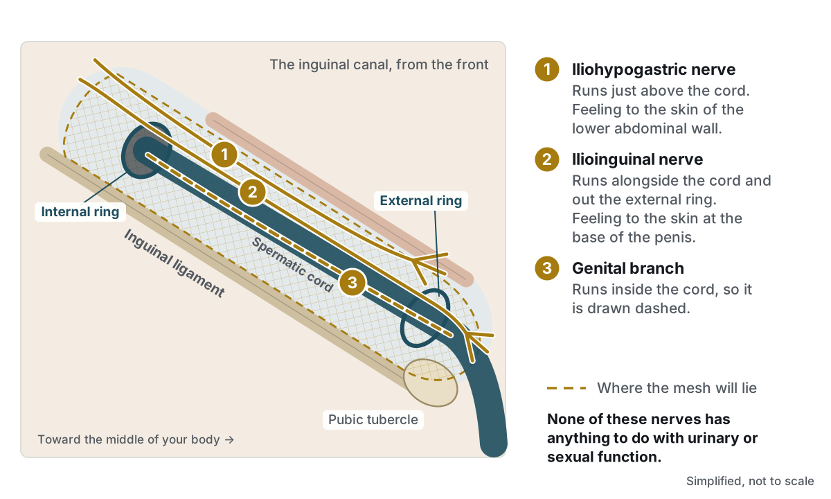
- Protecting the nerves: three nerves run through this area: the ilioinguinal, iliohypogastric, and genital branch of the genitofemoral nerve. The ilioinguinal and iliohypogastric nerves are identified early and protected throughout the case. The genital branch is not usually seen directly; the surgeon knows where it runs and takes steps to avoid it. Together, this lowers the risk of numbness or chronic pain afterward.
- Reducing the hernia: the bulging tissue (hernia sac) is gently freed from the surrounding structures and either returned to the abdomen or tied off and removed, depending on its size and location.
- Placing the mesh: a flat synthetic mesh is laid over the floor of the inguinal canal and secured in place, reinforcing the weak area without tension. The surgeon ensures the mesh lies flat to allow proper healing, with no folds or gaps that could let the hernia come back.
- Closing up: the layers of tissue are closed over the mesh, and the skin incision is closed with absorbable sutures placed deep to the skin and a waterproof skin glue (no staples or visible stitches to remove).
- Nerve block: a numbing medication is often injected to numb the groin nerves, reducing pain afterward and cutting down on the need for narcotics.
See it step by step: an animated guide to where the hernia comes from and how the mesh goes in ↓
Possible complications
- Bleeding or infection, lowered by careful cautery, antiseptic skin prep, sterile technique, and antibiotics before surgery
- Nerve injury or chronic groin pain, lowered by protecting the ilioinguinal and iliohypogastric nerves and by knowing and avoiding the path of the genital nerve during the repair
- Injury to the spermatic cord structures (in men) or (in women), or to nearby blood vessels. Uncommon, but possible given the close anatomy in this region
- Hernia recurrence, lowered by making sure the mesh lies completely flat with no folds
- Numbness around the incision, which is common and usually improves over time
Pain control after surgery
Pain is managed with a combination approach so you need fewer narcotics:
- NSAIDs, such as Advil (ibuprofen) or Celebrex (celecoxib), to reduce inflammation
- Tylenol (acetaminophen) for pain and fever
- Robaxin (methocarbamol), a muscle relaxant
- Narcotics only if needed, for the shortest time possible
Recovery: what to expect
- Hospital stay: most patients go home the same day, with mild pain, swelling, or bruising.
- Activity: walk as tolerated; avoid anything strenuous.
- Lifting: nothing over 10 pounds until your 2 week visit, when we reassess.
- Wound care: you can shower the day of surgery, but do not submerge your incision in a pool, tub, or other body of water for at least 4 weeks. The incision is closed underneath the skin and sealed with a waterproof glue.
- Sun protection: healing incisions sunburn easily and can discolor, sometimes permanently, so keep them covered.
For more information, see Recovery and getting back to life.
- Fever over 101°F
- Pain that medication doesn't control
- Redness, swelling, or drainage at an incision
- Trouble urinating or breathing
- Significant constipation
If it's after hours, your call will be routed to our answering service. The on-call surgeon or PA will receive your message and contact you.
For uncontrolled pain, shortness of breath, chest pain, or inability to keep fluids down, seek urgent medical attention right away rather than waiting for office hours.