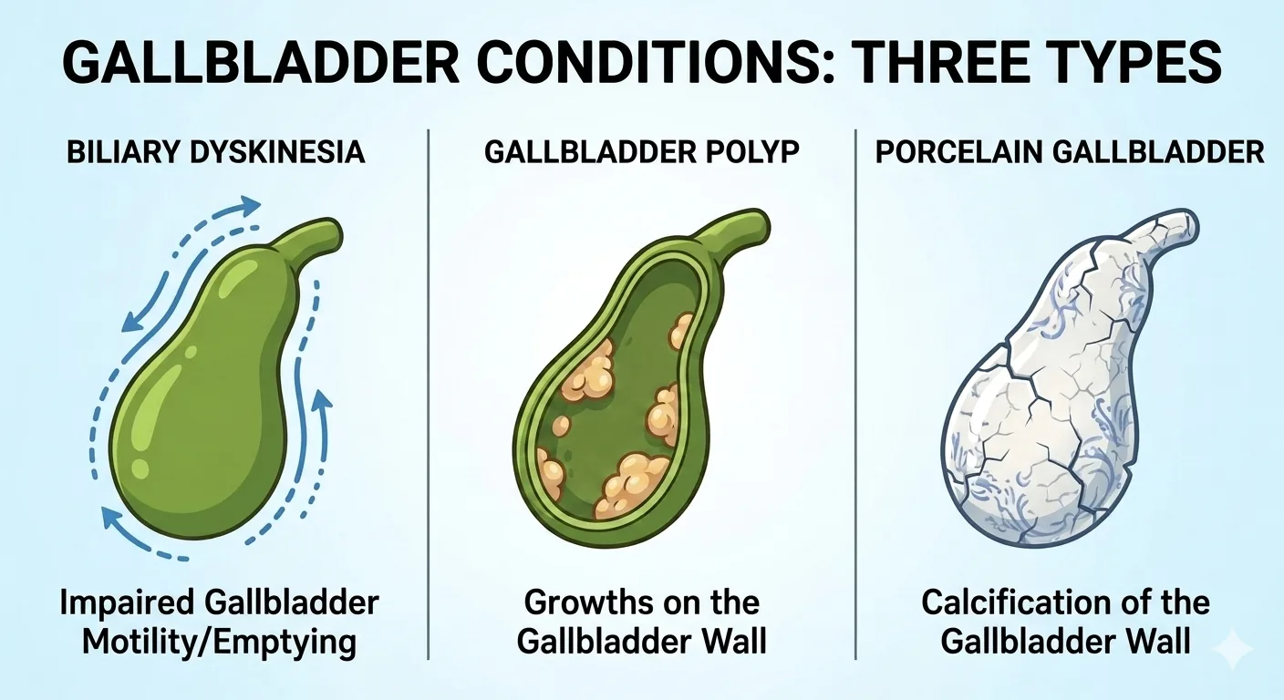
When you come to my office, most gallbladder conversations start with a stone on ultrasound. Three conditions do not, and none of them are common. In my practice, biliary dyskinesia accounts for roughly one in ten of the gallbladder patients I see. Polyps are fewer than one in a hundred. Porcelain gallbladder I see about once a year. Each one raises the same question in a different way: does this gallbladder need to come out?
When the Gallbladder Will Not Empty: Biliary Dyskinesia
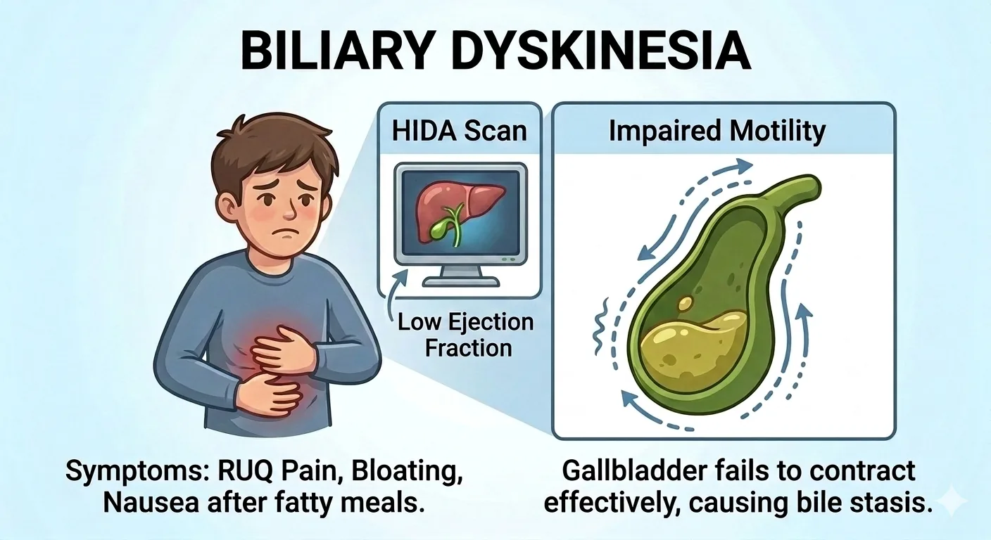
Some patients describe textbook biliary colic pain in the right upper quadrant after fatty meals, but their ultrasound is clean. No stones, no sludge, a normal-appearing gallbladder wall. When the pain is real but the imaging is silent, we look at function instead of anatomy.
We test this with a CCK-HIDA scan, the same hepatobiliary scintigraphy test introduced in Part 2, Physiology, this time using a synthetic form of cholecystokinin (CCK) to stimulate the gallbladder directly. The scan measures how much bile the gallbladder actually empties over 60 minutes, reported as a gallbladder ejection fraction (GBEF). At my institution we treat a GBEF below 35% as abnormal. That number is a local convention, not an agreed standard: the systematic review that anchors this literature found that centers used different cutoff values and that the studies were too weak methodologically to recommend one.¹ In appropriately selected patients with a low GBEF and no other explanation for their symptoms, cholecystectomy is an accepted treatment, even when the ultrasound shows no stones at all.²
The ejection fraction does not tell me everything. During the scan, the CCK injection sometimes reproduces the exact pain that brought the patient in. When that happens, I give it real weight in deciding whether to offer surgery. I apply the same reasoning at the other end of the range. Some gallbladders empty too fast, above 70%, which is termed a hyperkinetic gallbladder. More surgeons are offering cholecystectomy for these patients, and the review I cite here states that quality data supporting that practice do not yet exist.² I still offer surgery to selected hyperkinetic patients whose pain is reproduced during the scan, and I tell them where the evidence stands before we decide.
When Growths Appear on the Lining: Gallbladder Polyps
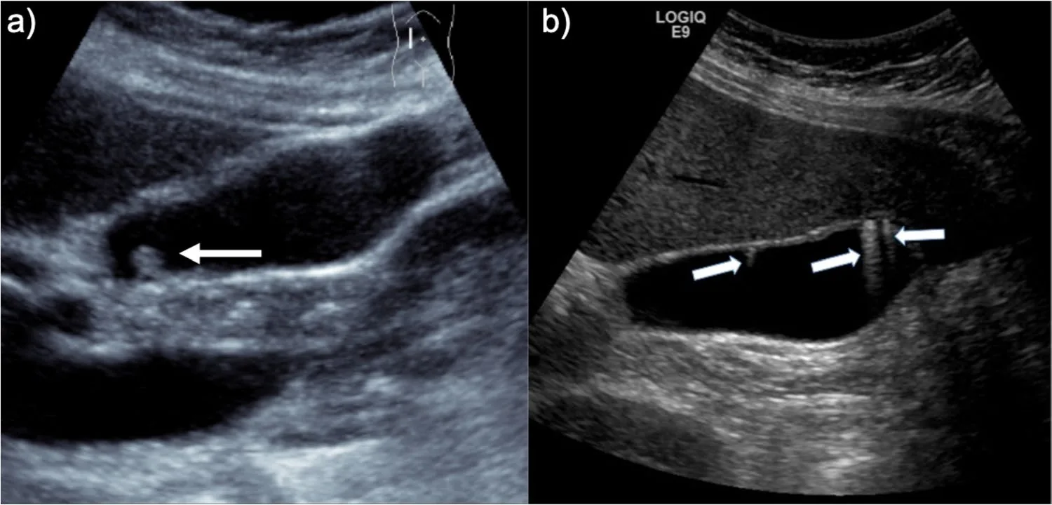
Polyps are growths that project from the gallbladder lining into the interior space. They can be seen on abdominal ultrasound, and the overwhelming majority are benign cholesterol polyps rather than true neoplasms.³
Telling a polyp from a stone comes down to a few features on the scan. A stone rolls when we reposition you on the exam table, and it casts a dark acoustic shadow behind it, shown on a real scan in Part 8, Imaging. A polyp does neither. It stays where it is, because it is attached to the wall, and it does not shadow.³ It may sit flat against the lining (sessile) or hang from a stalk (pedunculated). If the ultrasound shows a bright trailing streak behind the lesion, called a comet tail artifact, that points to a cholesterol deposit rather than a true polyp, and the guideline framework below does not apply to it.³ I will also put color Doppler on an unclear lesion: a polyp is living tissue and can show blood flow, while a stone has no blood supply at all.
The 2022 update to the joint ESGAR/EAES/EFISDS/ESGE guideline lays out a specific size and risk-factor framework:³
- **10 mm or larger:** cholecystectomy is recommended, if the patient is fit for and accepts surgery
- **6 to 9 mm with one or more risk factors** (age over 60, a history of primary sclerosing cholangitis, Asian ethnicity, or a sessile shape without a stalk, including focal wall thickening over 4 mm): cholecystectomy is recommended
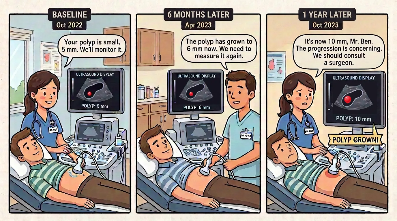
- **6 to 9 mm without risk factors, or 5 mm or smaller with risk factors:** follow-up ultrasound at 6 months, 1 year, and 2 years; surveillance stops if there is no growth by 2 years
- **5 mm or smaller without risk factors:** no follow-up needed
- **Growth to 10 mm during surveillance:** cholecystectomy is advised
- **Growth of 2 mm or more within the 2-year window:** re-evaluate size and risk factors together, often with a multidisciplinary discussion, to decide between continued monitoring and surgery
The age threshold moved from over 50 in the original 2017 guideline to over 60 in the 2022 update. That change was based on a systematic review of more than 5,400 polyps, which found that age over 60, not 50, was the threshold significantly associated with malignancy.³
When the Wall Calcifies: Porcelain Gallbladder
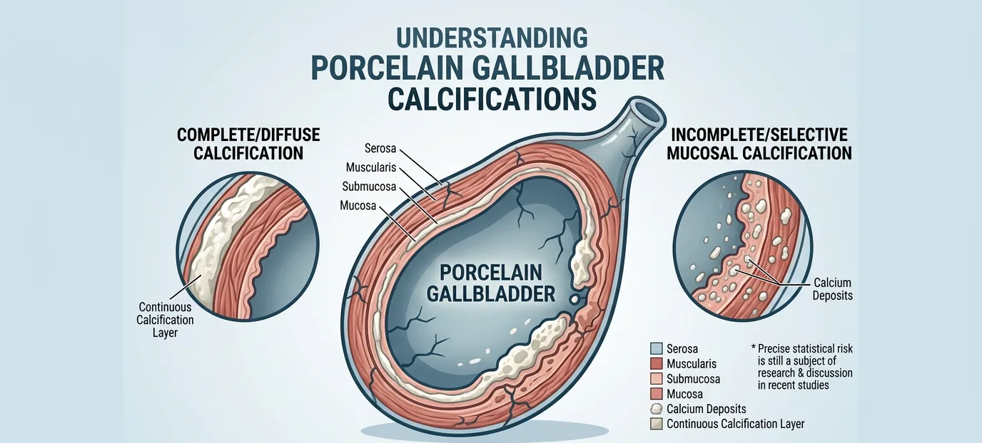
Porcelain gallbladder describes a gallbladder wall that has calcified after years of chronic, low-grade inflammation, giving it a hardened, chalky appearance on imaging that resembles porcelain.⁴ For decades, this finding was treated as an automatic indication for surgery because of reported cancer rates that ran from 12% to 33% in the older literature, with one 1967 series reporting 61.5%.⁴ More recent, better-controlled studies have found the true incidence of gallbladder cancer in porcelain gallbladder to be considerably lower than those older figures suggested.⁴
The pattern of calcification matters. A gallbladder with complete, uniform calcification throughout the wall carries a lower cancer risk than one with patchy, incomplete, or selective mucosal calcification, which has been associated with a meaningfully higher rate of malignancy.⁴ Given that risk, and in the absence of a reliable way to rule out early cancer without removing the organ, I generally recommend cholecystectomy for porcelain gallbladder, particularly when the calcification pattern is incomplete or the patient is otherwise a good surgical candidate.
The Common Thread
These three arrive in different ways. Biliary dyskinesia shows up as pain, with a normal ultrasound and nothing abnormal except the ejection fraction on the CCK-HIDA scan. Polyps and porcelain gallbladder are usually found by accident, on a scan ordered for something else, and neither one typically causes right upper quadrant pain. So one announces itself and two do not. The underlying question is still the same one we ask with every patient: does the risk of leaving this gallbladder in place outweigh the risk of taking it out? These three need a more individual answer than a stone does, which is why they deserve their own conversation before we move into how the surgery itself is performed.
For more on gallbladder disease and when removal is recommended, see my gallbladder patient guide. Missed the earlier posts in this series? You can find them all on the blog.
Up next: Part 4, Surgery, how the operation is actually done.
References
1. DiBaise JK, Oleynikov D. Does gallbladder ejection fraction predict outcome after cholecystectomy for suspected chronic acalculous gallbladder dysfunction? A systematic review. Am J Gastroenterol. 2003;98(12):2605-2611. https://doi.org/10.1111/j.1572-0241.2003.08772.x
2. Richmond BK. Biliary Dyskinesia, Controversies, Diagnosis, and Management: A Review. JAMA Surg. 2024;159(9):1079-1084. https://doi.org/10.1001/jamasurg.2024.0818
3. Foley KG, Lahaye MJ, Thoeni RF, et al. Management and follow-up of gallbladder polyps: updated joint guidelines between the ESGAR, EAES, EFISDS and ESGE. Eur Radiol. 2022;32:3358-3368. https://doi.org/10.1007/s00330-021-08384-w
4. Morimoto M, Matsuo T, Mori N. Management of Porcelain Gallbladder, Its Risk Factors, and Complications: A Review. Diagnostics (Basel). 2021;11(6):1073. https://doi.org/10.3390/diagnostics11061073