At the end of the last post, I said we would look at how we actually image the gallbladder. Here we go.
There are four tests: ultrasound, HIDA scan, MRCP, and CT. They are not four versions of the same test. Each one answers a different question, and which one you get depends on your symptoms.
So rather than walk through the tests one at a time, I am going to walk through three patients. Almost everyone with gallbladder pain arrives looking like one of them. These are a guide. Clinical decision making stays in the hands of the medical team who evaluates you.
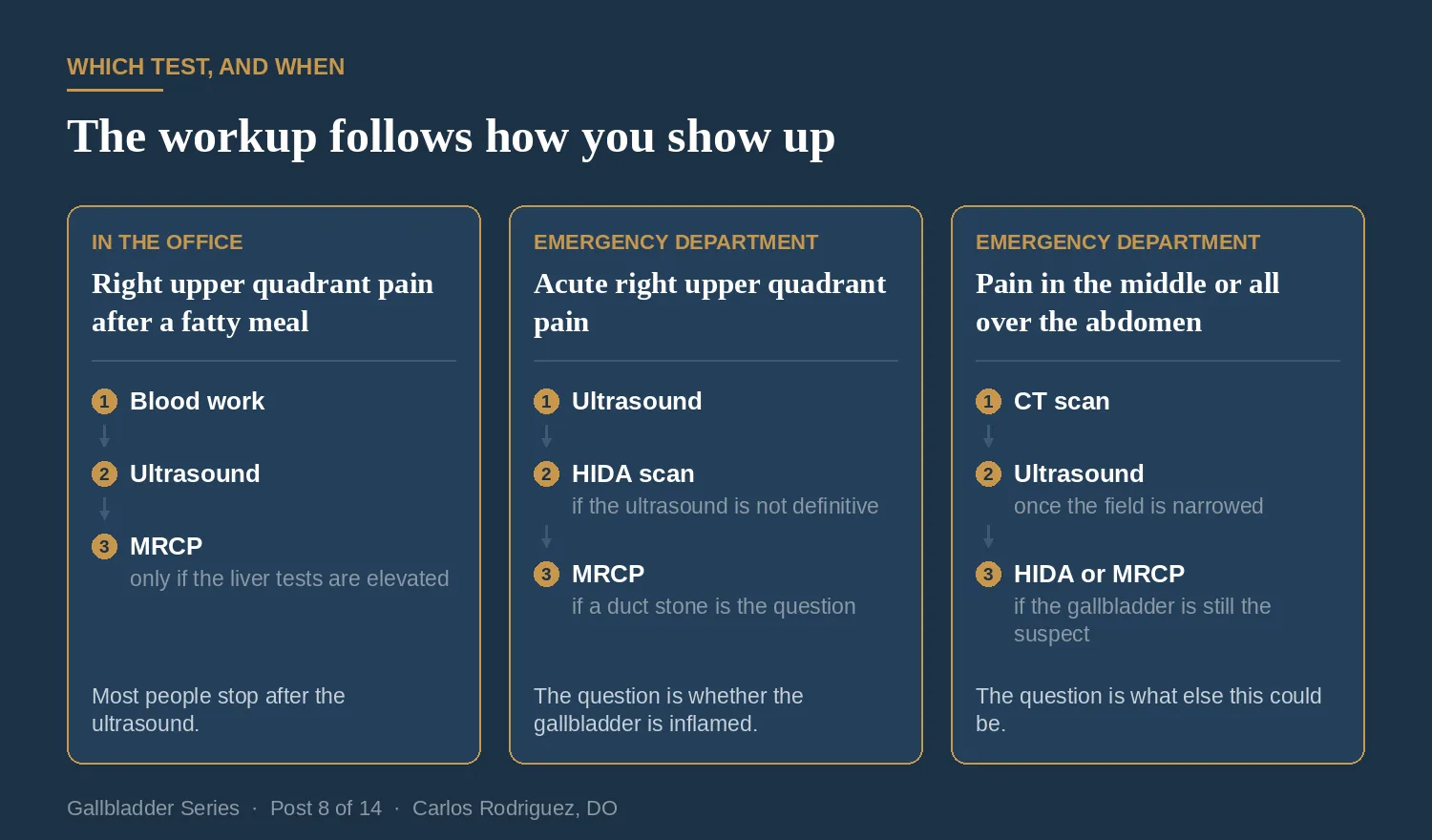
Patient One: Pain After a Fatty Meal, Seen in the Office
This is by far the most common situation I encounter. Pain in the right upper part of the abdomen that comes on after a heavy or greasy meal, lasts anywhere from thirty minutes to a few hours, and then goes away on its own. It may wake the patient at night. It may come with nausea. Between episodes the patient feels fine.
This patient is not sick in front of me. Nothing is urgent. The workup is two things: blood work and an ultrasound.
The Blood Work
A basic panel that includes liver enzymes and bilirubin. With this patient presentation these labs are usually normal, and normal is useful information. It tells me the bile is still draining.
The reason I check is that abnormal liver enzymes change the plan, and I will come back to that below.
The Ultrasound
Ultrasound is the first imaging test in nearly every case of suspected gallbladder disease. The Tokyo Guidelines 2018 recommend it as the first-choice imaging method¹ (p.43). Why? It is widely available, it is not invasive, it is easy to use, and it costs less than the alternatives. Ultrasound also does not involve radiation, contrast, or needles.

An ultrasound probe sends sound waves into the body and listens for the echoes, meaning the sound bouncing back. Tissues that reflect sound strongly show up bright. Fluid, which reflects almost nothing, shows up black.
A gallstone is dense. It bounces back nearly all of the sound that hits it, which means two things happen. The stone itself appears as a bright white spot, and the area directly behind it appears as a dark stripe, because almost no sound goes through the stone to reach the tissue beyond it. That dark stripe is termed an acoustic shadow.
The shadow matters more than the bright spot. Plenty of things inside a gallbladder can look bright. Only a stone reliably casts a shadow behind itself. A radiologist looking for stones is looking for three features together: bright spots, shadows behind them, and movement when the patient rolls onto their side².
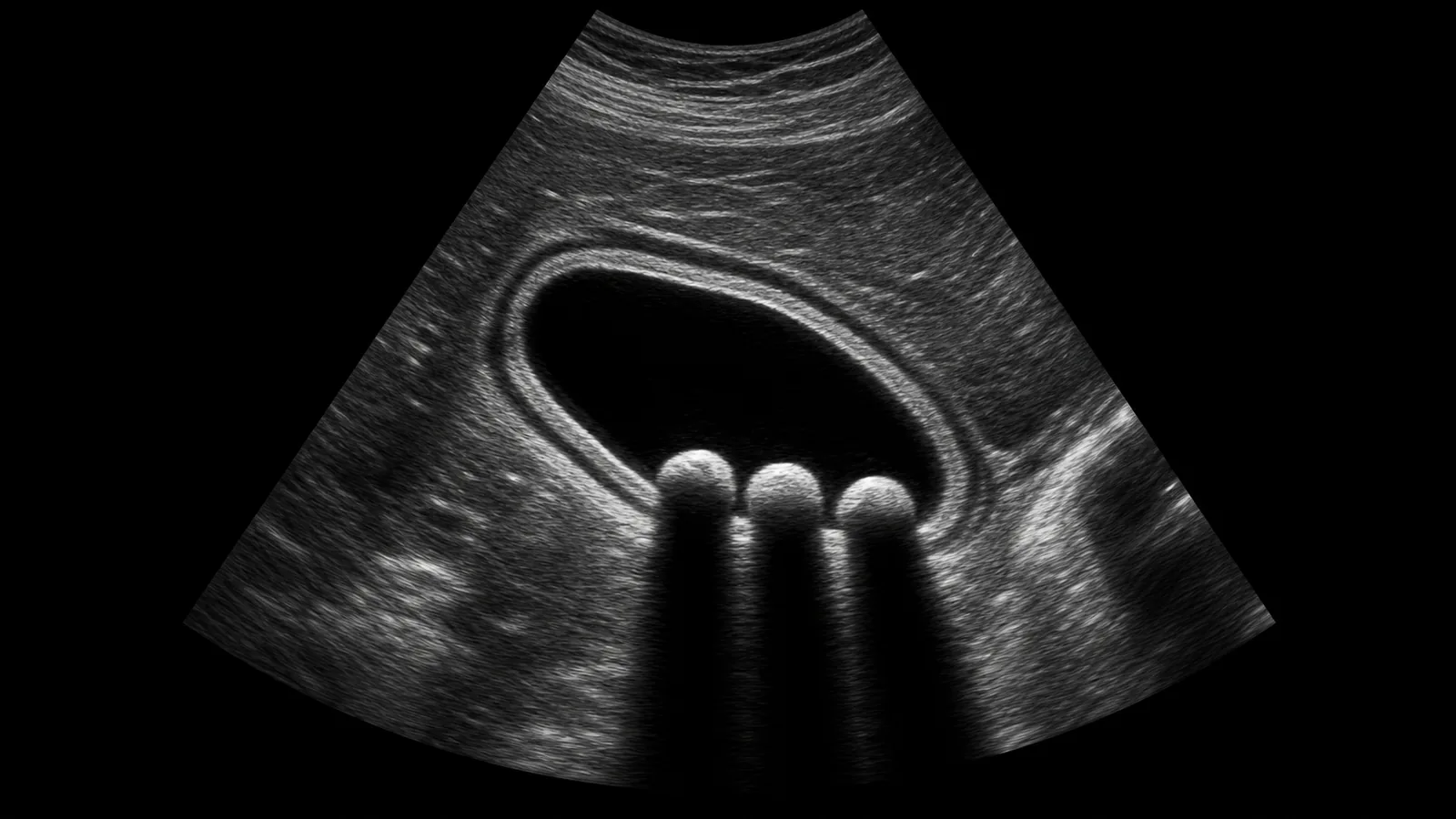
For this patient, that is the whole workup. Stones on the ultrasound plus a story of pain after fatty meals is enough to make the diagnosis and enough to offer surgery. No further imaging is needed.
Finding stones is not the same as finding disease. Most people with gallstones never develop symptoms, which I covered in Post 4 of this series, published July 27, 2026. What makes this patient a surgical candidate is the combination: stones on the picture and symptoms that match.
When the Liver Enzymes Are Elevated
Now change one thing. Same patient, same story, but the liver enzymes come back elevated.
That raises a different question. It suggests a stone may have left the gallbladder and lodged in the common bile duct, which is the tube carrying bile from the liver down to the intestine. That is a different problem with a different treatment, and I covered it in Post 6, published August 10, 2026.
The trouble is that ultrasound is not good at answering it. Ultrasound sees the gallbladder well and it can measure the diameter of the common bile duct, but it cannot see the whole duct. The duct sits deeper, and gas in the duodenum sits right in front of the part where stones tend to lodge.
A Cochrane review pooled five studies and 523 patients on this exact question. For finding a stone in the common bile duct, ultrasound was 73% sensitive, with a confidence interval running from 44% to 90%³. That range includes performance no better than a coin flip. The review puts it in patient terms: out of 100 people with a duct stone, about 73 are found and 27 are missed. The authors' conclusion is that many people may have common bile duct stones in spite of having a negative ultrasound or negative liver tests³.
So the ultrasound cannot answer it. This is where clinical judgment comes in, because there are two reasonable next steps: an MRCP, followed by ERCP if a stone is seen, or surgery with an intraoperative cholangiogram. Both are worth walking through.
MRCP: A Road Map of the Ducts
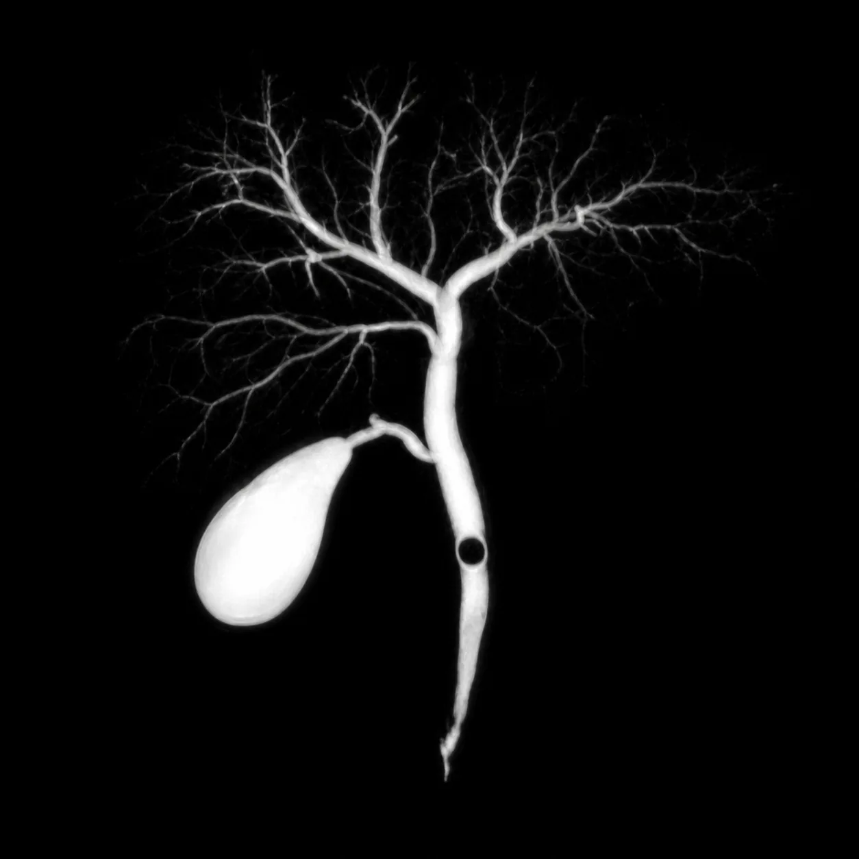
MRCP stands for magnetic resonance cholangiopancreatography. It is a specific set of MRI sequences tuned so that slow-moving fluid appears bright and everything else appears dark. Bile is fluid. On MRCP the entire bile duct system lights up like a road map, and a stone sitting inside it appears as a dark gap in the bright line.
Two things make this test useful. There is no radiation, and nothing is injected into the ducts. The bile already there works as the contrast.
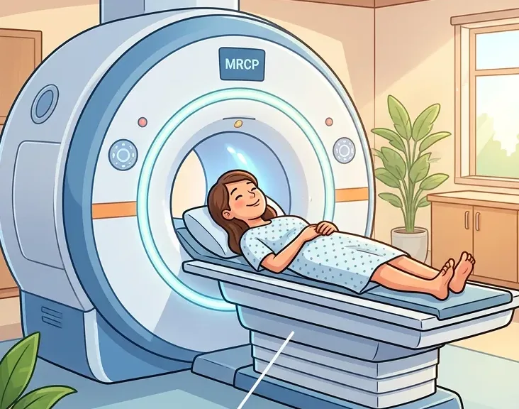
For the question of whether there is a stone in the common bile duct, MRCP is 93% sensitive and 96% specific, pooled across seven studies and 996 patients⁴. Compare that to 73% for ultrasound on the same question, from a Cochrane review published the same year³.
Both Cochrane reviews carry a caveat. Their evidence is current to 2012, and the included studies were of poor methodological quality by the reviewers' own assessment³ ⁴. MRCP hardware has improved since then, so the exact percentages may be different today.
The Other Option: Look During Surgery
There is a second way to answer the duct question, and it does not involve another scan. During the operation, dye can be injected into the duct and an X-ray taken on the spot. This is an intraoperative cholangiogram.
It answers the same question as the MRCP, it happens while the patient is already asleep for the gallbladder removal, and it does not add a separate appointment. I am going to leave the details there, because it belongs with the operation itself and that is Post 11 in this series.
The choice between the two comes down to how likely a duct stone is and what the surgeon and the hospital are set up to do. For an outpatient who is otherwise well, the MRCP can be scheduled and the timing is rarely the deciding factor. In the hospital it is a different calculation, and I come back to it with the next patient.
Patient Two: Acute Right Upper Quadrant Pain in the Emergency Department
Different patient. This one is not between episodes. The pain started hours ago, it has not stopped, it hurts to take a deep breath, and there may be a fever.
The question here is not whether there are stones. It is whether the gallbladder is inflamed right now.
The first test is still an ultrasound, for the same reasons as before, plus one more: it can be done at the bedside in the emergency department.
What the Ultrasound Is Looking for Now
The Tokyo Guidelines list the findings that point to acute cholecystitis: a gallbladder wall 4 mm or thicker, a gallbladder enlarged to 8 cm or more in its long axis or 4 cm in its short axis, stones or retained debris inside it, fluid collecting around the outside of it, and linear shadows in the fat surrounding it¹ (p.45). I walked through the Tokyo Guidelines and how they grade severity in Post 5, published August 3, 2026.
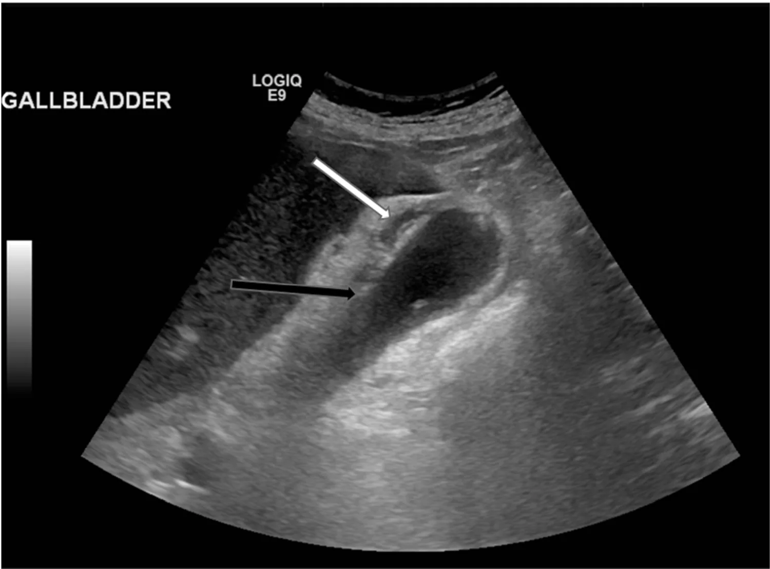
There is one more finding, and it is the only one that requires the patient to be awake and participating. The sonographer presses the probe directly over the gallbladder and asks the patient to take a deep breath. If the patient stops the breath because of pain under the probe, that is a positive sonographic Murphy sign. Murphy's sign was first described in 1982, in a study of 427 patients, and it is 63% sensitive and 93.6% specific⁶. It misses about a third of cases, so a negative sign does not rule out cholecystitis. A positive one is meaningful.
When the Ultrasound Is Not Definitive
Ultrasound has two limitations.
The first is that the test is operator and body dependent. Bowel gas can sit in front of the structures we want to see and obscure them. Body habitus changes how deep the sound must travel, which makes some structures hard to see clearly. In the largest meta-analysis of imaging in acute cholecystitis, 57 studies and 5,859 patients, ultrasound came out 81% sensitive and 83% specific⁷. Roughly one in five inflamed gallbladders is missed.
The second limitation is the common bile duct problem from Patient One.
When the ultrasound is equivocal and the patient still appears to have a gallbladder related illness, the next test is a HIDA scan, an MRCP, or both, depending on the clinical situation. If the question is whether the gallbladder is inflamed, that is the HIDA scan. If the question is whether there is a stone in the duct, that is the MRCP.
HIDA: The Test That Asks Whether the Gallbladder Works
Every other test on this list looks at structure. A HIDA scan looks at function.
HIDA stands for hepatobiliary iminodiacetic acid. A small amount of a radioactive tracer is injected into a vein. The liver pulls that tracer out of the blood and excretes it into bile, exactly the way it handles bile normally. A camera then watches where the bile goes over the next hour or two.
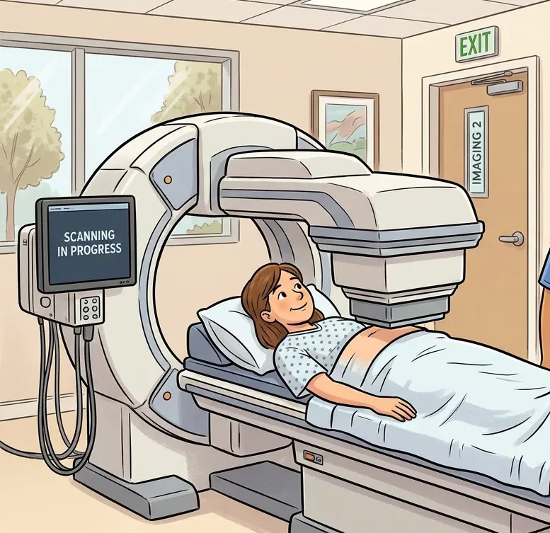
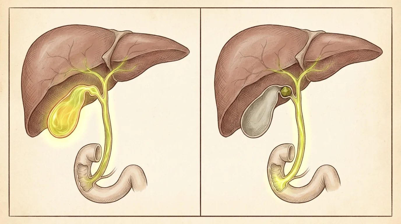
In a normal scan, tracer flows from the liver, fills the gallbladder, and continues down the bile duct into the intestine.
If the cystic duct, which is the gallbladder's only outlet, is blocked by a stone, the gallbladder never fills. The liver is still working, and the tracer still runs down the bile duct into the intestine. The spot where the gallbladder should appear stays blank.
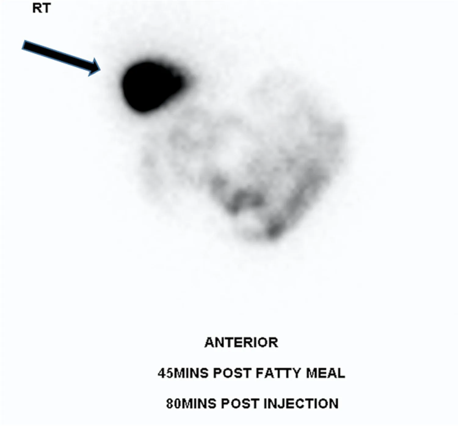
This is the most sensitive test we have for acute cholecystitis. In the Kiewiet meta-analysis, 57 studies and 5,859 patients, HIDA was 96% sensitive against 81% for ultrasound, and the difference was statistically significant⁷.
So why is ultrasound still the first test? The Tokyo Guidelines address this. They acknowledge that every study comparing the two has found HIDA to be the better test, and they still recommend ultrasound first, for cost and availability¹ (p.43). A HIDA scan takes one to two hours, requires nuclear medicine staffing, and is not available at every hospital at 2 a.m.
Two peer-reviewed meta-analyses do not agree on how specific a HIDA scan is. A 2012 analysis put HIDA specificity at 90%⁷. A 2024 meta-analysis of 22 studies put it at 63%, with pooled sensitivity of 91%⁸. Both are pooled estimates from the published literature. They used different inclusion criteria and drew on partly different studies.
What both agree on is that HIDA is the most sensitive test for acute cholecystitis. Where they differ is on how often it calls a healthy gallbladder abnormal. A false positive on a HIDA scan is not a trivial thing, because it can send someone to the operating room. That is why the scan gets interpreted alongside the patient's history, physical exam, and laboratory results, and never alone.
A HIDA scan is also used to evaluate biliary dyskinesia, which is covered in next week's post.
When the Wait Changes the Plan
MRCP is the most accurate non-invasive test for a duct stone. In the hospital it is also the one you wait for, and that wait can change what we do.
In a study of 100 consecutive patients admitted with gallstone pancreatitis at one hospital, the median wait was 4 days for an MRCP, against 1 day for an ultrasound and same day for a CT⁹. A separate review of 234 inpatient MRCPs at another hospital found a median of 2 days from request to scan, and then a median of 1 more day to get the scan read, with the longest waits running to two weeks¹⁰.
An MRI takes a while to perform and demand for MRI time is high, so the wait reflects how a hospital is staffed and scheduled. The authors of the second study say the fix is faster access and faster reporting rather than ordering fewer scans¹⁰.
Here is what it means in practice. If the wait for an MRCP is long enough that the patient would sit in the hospital for it, we go to the operating room instead: cholecystectomy with an intraoperative cholangiogram. The operation had to happen anyway, and the cholangiogram answers the duct question while the patient is already asleep.
Studies comparing an MRCP-first pathway against going to surgery with an intraoperative cholangiogram have generally found shorter stays in the surgery-first group. The one randomized trial built to measure it, 122 patients across five hospitals, estimated 1.2 extra days with MRCP, but that difference did not reach statistical significance (p=0.0799), the median stay was 4 days in both groups, and the authors concluded the two pathways were equivalent¹¹. I am not going to quote a number of extra hospital days as though it were known, because it is not.
Who Needs a Duct Study and What Happens Next
Not everyone with gallstones and abnormal labs needs an MRCP. The American Society for Gastrointestinal Endoscopy sorts patients by how likely a duct stone is¹².
Three findings put someone at high risk, and those patients may go straight to ERCP rather than to another scan:
- A stone visible in the duct on ultrasound or other imaging.
- A total bilirubin above 4 mg/dL together with a dilated bile duct.
- Ascending cholangitis, meaning an infected, obstructed bile duct. Not every case of cholangitis goes to ERCP right away. I covered the timing in Post 6, published August 10, 2026.
Intermediate risk is where an MRCP earns a place: abnormal liver blood tests, age over 55, or a dilated duct without the other findings. The guideline suggests MRCP or endoscopic ultrasound for those patients, and it estimates their chance of having a duct stone at 10% to 50%¹². A dilated duct means wider than 6 mm with the gallbladder still in place, or wider than 8 mm after it has been removed. Patients with none of those findings do not need a duct study at all.
Two rules follow from all of this, and they are the ones I actually use:
- If a stone is seen on the MRCP, the patient goes to ERCP first, so the stone comes out before the gallbladder does.
- If a stone is seen on the intraoperative cholangiogram and it cannot be cleared in the operating room, the patient goes to ERCP after surgery.
Either way the duct gets cleared. What changes is the order.
Patient Three: Pain in the Middle or All Over the Abdomen
Third patient, also in the emergency department, but the pain is not localized to the right upper quadrant. It is in the middle of the abdomen, or the patient cannot point to one spot, or it hurts everywhere. There may be vomiting.
Now the gallbladder is one item on a list. So is pancreatitis, a perforated ulcer, a bowel obstruction, and appendicitis, among others. Ordering an ultrasound first means answering one question about one organ when the question on the table is much broader.
This patient gets a CT scan.
A CT of the abdomen and pelvis covers the whole field in about ten seconds and answers a different question: what else could this be. It sees the liver, pancreas, bowel, appendix and kidneys in the same picture.
What CT Shows of the Gallbladder
CT is capable of showing an inflamed gallbladder.
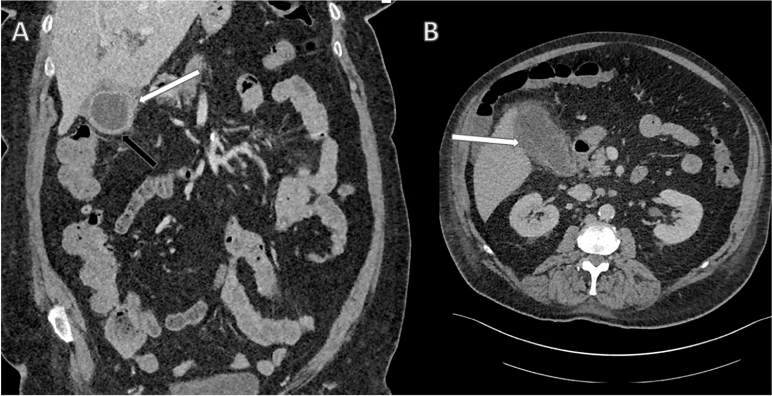
CT is also where we look for the complications. The Tokyo Guidelines recommend contrast-enhanced CT or contrast-enhanced MRI when gangrenous cholecystitis is suspected, meaning the gallbladder wall has begun to die¹ (p.48). One specific finding, the interrupted rim sign, where the contrast-enhanced wall shows a break in its bright rim, is 73% sensitive with a 95% negative predictive value¹ (p.49). Those findings are underestimated on ultrasound.
Where CT Falls Short
CT is much less reliable at finding the stones themselves, and the reason is density.
A stone made largely of calcium is denser than the bile around it, so it shows up bright and obvious. A stone made largely of cholesterol can be almost exactly as dense as bile, which makes it invisible. The authors of the imaging review say that a significant proportion of stones "are isoattenuating relative to surrounding bile and may be radiologically occult on CT"⁵.
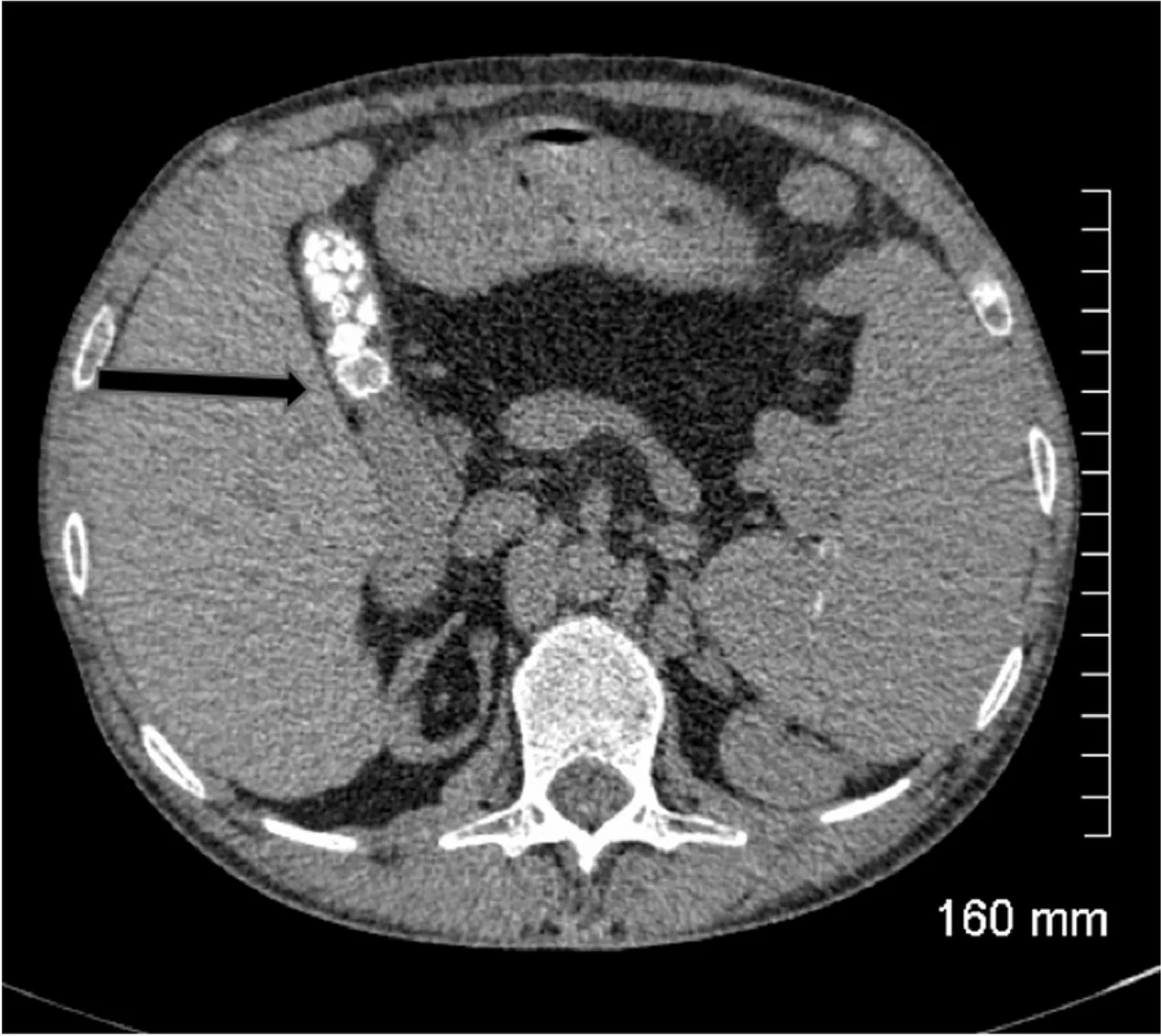
That is a large part of why a CT reported as showing no gallstones does not mean there are none. The same review states: "CT remains inferior to ultrasound at assessing the gallbladder and results in a significant radiation dose to the patient. As such, ultrasound is the imaging modality of choice for initial assessment of suspected gallbladder pathology. CT however can be very effective at assessing extra-biliary gallstone pathology and complications arising from gallstone pancreatitis and cholecystitis"⁵.
So this patient often ends up getting an ultrasound too, after the CT has narrowed the field. That is not duplication. The CT answered what else this could be. The ultrasound answers what the gallbladder is doing.
An Update Worth Making
For years the standard line on CT for acute cholecystitis was that it had not been studied properly. The 2012 meta-analysis found only one CT study that met its inclusion criteria, and that single study reported 94% sensitivity with 59% specificity⁷. Those numbers got quoted for a decade.
That has changed.
Two meta-analyses published in 2024 pooled CT data. One analyzed 64 studies and compared CT against ultrasound in the same patients, finding CT 83.9% sensitive and 94% specific, against 79.0% and 93.6% for ultrasound. Neither difference was statistically significant, with p values of 0.44 and 0.85¹³. The other pooled 22 studies and put CT at 78% sensitive and 81% specific⁸.
The current reading is that CT and ultrasound perform comparably for making the diagnosis itself. Ultrasound remains the first test, for cost, availability and radiation. CT is valuable when the diagnosis is uncertain or a complication is suspected.
The Radiation Question
Patients do ask about this.
A CT of the abdomen and pelvis delivers an effective dose of about 7.7 mSv, which is roughly what a person in the United States absorbs from natural background radiation over 2.6 years¹⁴. Ultrasound and MRI use none¹⁴.
That is not a reason to refuse a CT when it is the right test. A missed perforated gallbladder is a far larger risk than 7.7 mSv. It is a reason not to order one reflexively when an ultrasound would answer the question.
What About ERCP?
ERCP comes up in every conversation about biliary imaging, so it belongs here even though it is not really an imaging test.
ERCP is a procedure. A scope goes down through the mouth to the point where the bile duct empties into the intestine, dye is injected up into the duct, and images are taken. The same scope can remove the stone during the same procedure.
That is why the guidelines route high-risk patients to ERCP instead of to an MRCP first¹². If a stone is almost certainly there, there is no reason to take a picture of it and then schedule a second procedure to remove it.
I covered ERCP in detail in Post 6, published August 10, 2026, so I will leave it there.
Required Reading
Four of the images in this post come from one paper.
Murphy and colleagues, writing in Insights into Imaging in 2020, published "Gallstones top to toe: what the radiologist needs to know"⁵. It is the best single primer on gallbladder imaging I have found.
It walks the reader through the entire biliary tree from the gallbladder to the duodenum, and at every stop it shows you the actual images: what a stone looks like on ultrasound, on plain film, on CT, on MRI, on cholescintigraphy, on ERCP. It covers acute and chronic cholecystitis, gangrenous and emphysematous disease, porcelain gallbladder, Mirizzi syndrome, gallstone ileus, Bouveret syndrome, and the complications that follow cholecystectomy. It is open access. No paywall, no login.
If you take care of patients with gallbladder disease, read it.
The Part Patients Should Take Away
If you get sent for a second scan, it is not because the first one failed. It is because the first one answered its question and a different question remains unanswered.
Ultrasound tells us about stones and inflammation. A HIDA scan tells us whether your gallbladder still works. MRCP maps your bile ducts and looks for a stone in them. CT looks at everything around the gallbladder and finds the complications.
No single test sees all four things. That is not a gap in the technology. It is why we have four tests.
For more on gallbladder disease and when removal is recommended, see my gallbladder patient guide. Missed the earlier posts in this series? You can find them all on the blog.
Up next: the three gallbladder problems that are not caused by a stone at all, and what to do about each of them.
References
1. Yokoe M, Hata J, Takada T, et al. Tokyo Guidelines 2018: diagnostic criteria and severity grading of acute cholecystitis (with videos). J Hepatobiliary Pancreat Sci. 2018;25(1):41-54. https://doi.org/10.1002/jhbp.515
2. Yu MH, Kim YJ, Park HS, Jung SI. Benign gallbladder diseases: Imaging techniques and tips for differentiating with malignant gallbladder diseases. World J Gastroenterol. 2020;26(22):2967-2986. https://doi.org/10.3748/wjg.v26.i22.2967
3. Gurusamy KS, Giljaca V, Takwoingi Y, et al. Ultrasound versus liver function tests for diagnosis of common bile duct stones. Cochrane Database Syst Rev. 2015;(2):CD011548. https://doi.org/10.1002/14651858.CD011548
4. Giljaca V, Gurusamy KS, Takwoingi Y, et al. Endoscopic ultrasound versus magnetic resonance cholangiopancreatography for common bile duct stones. Cochrane Database Syst Rev. 2015;(2):CD011549. https://doi.org/10.1002/14651858.CD011549
5. Murphy MC, Gibney B, Gillespie C, Hynes J, Bolster F. Gallstones top to toe: what the radiologist needs to know. Insights into Imaging. 2020;11(1):13. https://doi.org/10.1186/s13244-019-0825-4
6. Ralls PW, Halls J, Lapin SA, Quinn MF, Morris UL, Boswell W. Prospective evaluation of the sonographic Murphy sign in suspected acute cholecystitis. J Clin Ultrasound. 1982;10(3):113-115. https://doi.org/10.1002/jcu.1870100305
7. Kiewiet JJS, Leeuwenburgh MMN, Bipat S, Bossuyt PMM, Stoker J, Boermeester MA. A systematic review and meta-analysis of diagnostic performance of imaging in acute cholecystitis. Radiology. 2012;264(3):708-720. https://doi.org/10.1148/radiol.12111561
8. Childs DD, Lalwani N, Craven T, et al. A meta-analysis of the performance of ultrasound, hepatobiliary scintigraphy, CT and MRI in the diagnosis of acute cholecystitis. Abdom Radiol (NY). 2024;49(2):384-398. https://doi.org/10.1007/s00261-023-04059-w
9. Chen SE, Sokhal BS, Nnaji M, et al. Acute Gallstone Pancreatitis: If a Picture Is Worth a Thousand Words, How Many Images Do We Need? Cureus. 2023;15(1):e33666. https://doi.org/10.7759/cureus.33666
10. Milburn JA, Hall S, Sinha S, Ramsingh J, Ravindran R. Inpatient magnetic resonance cholangiopancreatography: does it increase the efficiency in emergency hepatopancreaticobiliary surgery services? Ann R Coll Surg Engl. 2017;99(4):289-294. https://doi.org/10.1308/rcsann.2016.0291
11. Staubli SM, Kettelhack C, Oertli D, et al. Efficacy of intraoperative cholangiography versus preoperative magnetic resonance cholangiography in patients with intermediate risk for common bile duct stones. HPB (Oxford). 2022;24(11):1898-1906. https://doi.org/10.1016/j.hpb.2022.05.1346
12. Buxbaum JL, Abbas Fehmi SM, Sultan S, et al. ASGE guideline on the role of endoscopy in the evaluation and management of choledocholithiasis. Gastrointest Endosc. 2019;89(6):1075-1105.e15. https://doi.org/10.1016/j.gie.2018.10.001
13. de Oliveira GS, Torri GB, Gandolfi FE, et al. Computed tomography versus ultrasound for the diagnosis of acute cholecystitis: a systematic review and meta-analysis. Eur Radiol. 2024;34(11):6967-6979. https://doi.org/10.1007/s00330-024-10783-8
14. Radiological Society of North America and American College of Radiology. Patient Safety: Radiation Dose in X-Ray and CT Exams. RadiologyInfo.org. https://www.radiologyinfo.org/en/info/safety-xray (accessed 8/23/2026; last site update 4/15/2025)
Figures 6, 9, 11 and 12 are reproduced from reference 5, Murphy MC et al., Insights into Imaging 2020;11(1):13, which is published under a Creative Commons Attribution 4.0 International License (CC BY 4.0), https://creativecommons.org/licenses/by/4.0/. They are Figures 9, 6, 10 and 4 of the original article respectively, cropped from the article page and otherwise unaltered. Their quoted captions are the original captions as printed.
Page numbers cited for reference 1 are the printed page numbers in Journal of Hepato-Biliary-Pancreatic Sciences volume 25, read from the published PDFs. Recommendation numbers quoted from that guideline refer to its own numbered statements: ultrasound as first-choice imaging is Recommendation 1, level C (p.43); contrast-enhanced CT or MRI for gangrenous cholecystitis is Recommendation 2, level C (p.48).