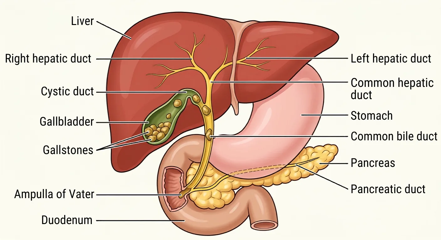
In the last post, we covered acute cholecystitis: when a stone stays wedged in the neck of the gallbladder or the cystic duct, seals it off, and the gallbladder becomes inflamed and/or infected. Sometimes a stone slips out of the cystic duct and drops into the common bile duct. When that happens, the problem stops being about the gallbladder alone and starts being about the drainage of the whole liver, especially if the stone becomes lodged in the common bile duct. This is termed choledocholithiasis.
What Is Choledocholithiasis?
The common bile duct forms where two smaller ducts meet: the common hepatic duct, which drains bile out of the liver, and the cystic duct, which drains the gallbladder. From that junction, the common bile duct carries bile the rest of the way to the small intestine (duodenum). When a stone lodges in that shared channel instead of staying up in the gallbladder, it blocks the outflow for the entire biliary system, not just the gallbladder. This is choledocholithiasis, which simply means a stone in the common bile duct.¹
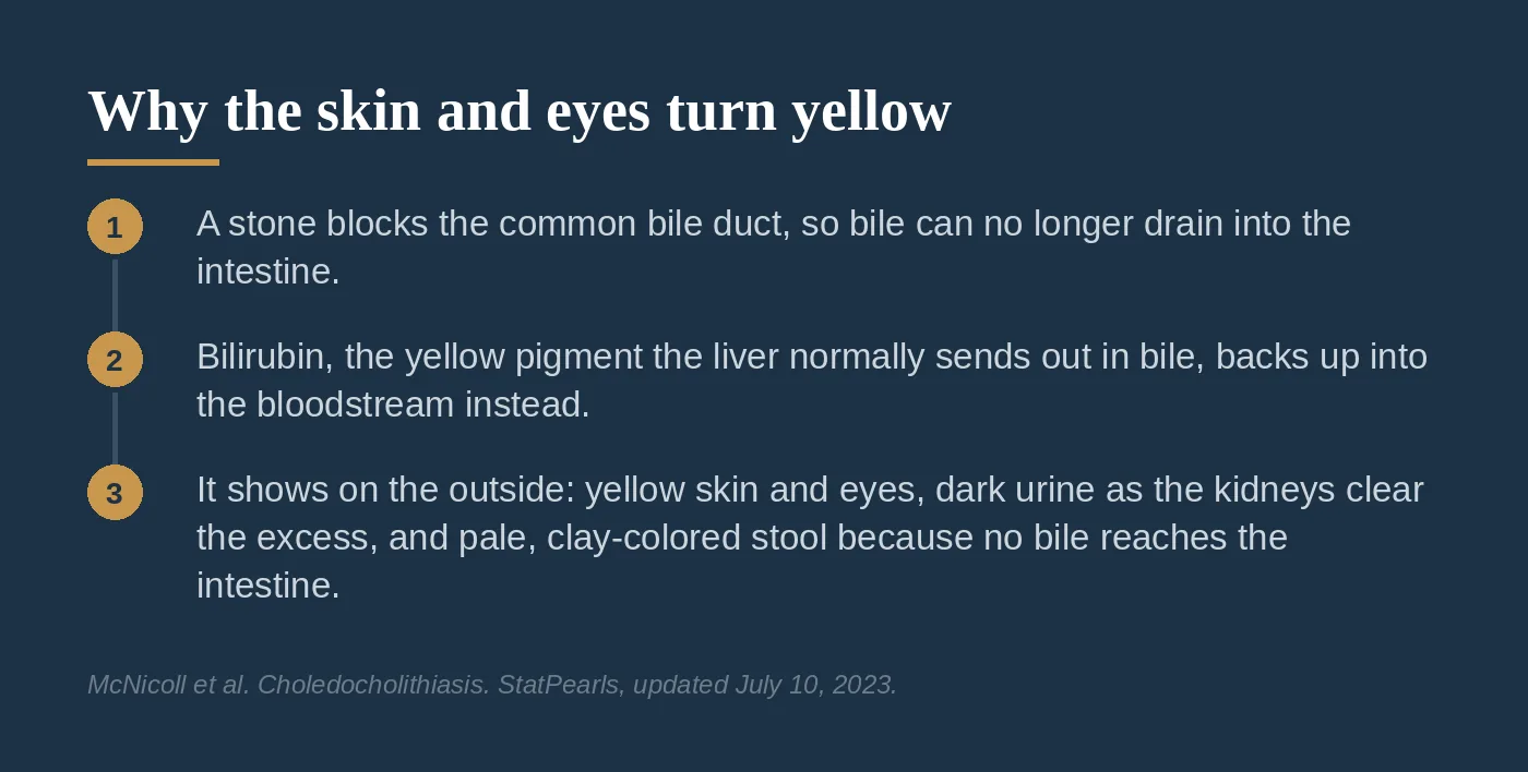
Because the duct is blocked, bilirubin, the yellow pigment the liver normally clears from the body by sending it out in bile, has nowhere to go. It backs up into the bloodstream instead of draining into the intestine. Blood levels climb, and the body starts to show it.
Recognizing the Blockage
A backed-up bile duct produces a recognizable pattern:
- Yellowing of the skin, the whites of the eyes, and the area under the tongue, as bilirubin builds up in the blood
- Dark urine, because the kidneys start clearing the excess bilirubin that the bile duct cannot
- Pale or clay-colored stool, because bile never reaches the intestine to give stool its normal brown color
We confirm the diagnosis with blood work and imaging. The blood work shows elevated bilirubin and elevated liver enzymes; a total bilirubin above 3 to 4 mg/dL is a strong pointer toward a stone in the duct.¹ On imaging, a standard abdominal ultrasound is very good at showing that the common bile duct is dilated, right about 90% of the time, but it actually sees the stone itself in only 15% to 40% of cases, because the lower end of the duct is hard to image through overlying bowel gas.¹ When the ultrasound is not conclusive, the next study is usually MRCP, a form of MRI that can map the bile ducts without contrast. MRCP is about 92% sensitive and close to 100% specific for a stone in the common bile duct.¹
How We Clear the Stone
There are two main ways to remove a stone from the common bile duct.
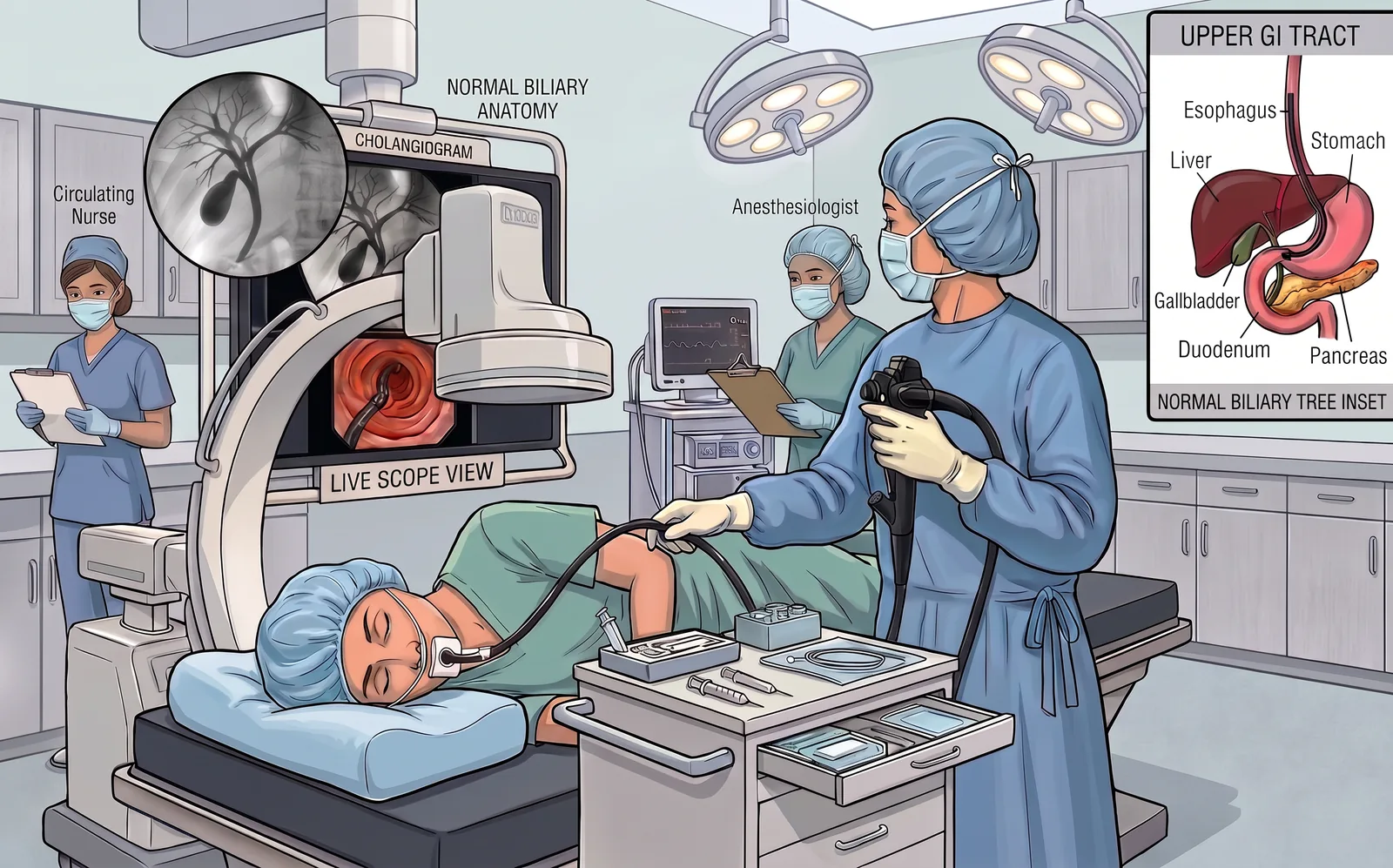
The first is ERCP, which stands for endoscopic retrograde cholangiopancreatography. A thin scope is passed through the mouth and stomach and down to the sphincter of Oddi at the ampulla of Vater, the shared opening we covered in Part 2, where bile and pancreatic juice both enter the intestine. A small cut in that muscular opening, called a sphincterotomy, widens it, and the stone is pulled out directly through the opening with a snare, a basket, or a balloon.¹
The second option is a common bile duct exploration, done at the time of the gallbladder operation. The surgeon opens or accesses the duct during the cholecystectomy and clears the stone, often with the same minimally invasive techniques used to remove the gallbladder.¹ Which route is chosen depends on the patient, the anatomy, when the stone is found, and the resources of the hospital.
When Infection Sets In: Cholangitis
A blocked duct creates stagnant bile, and stagnant bile is an easy target for bacteria. When the bile trapped behind a blockage becomes infected, that is called cholangitis, and it is a distinct and far more dangerous problem than a duct stone not causing any problem.³
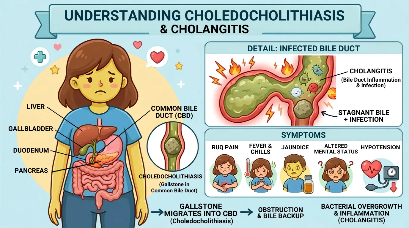
Two named patterns help gauge how serious it is:
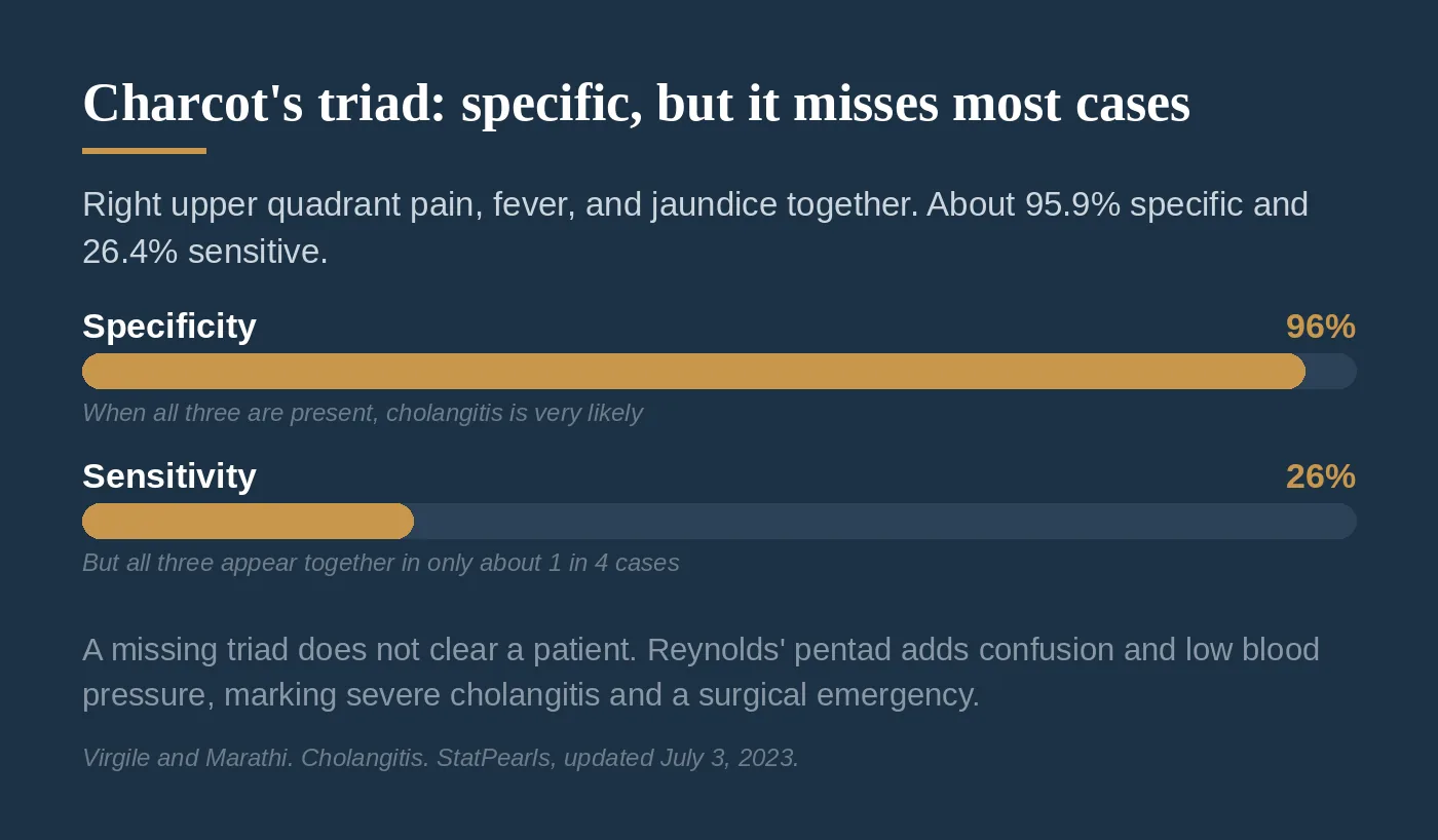
- Charcot's triad: right upper quadrant pain, fever, and jaundice occurring together. This combination is very specific for cholangitis, about 95.9%, but it is present in only about 26.4% of cases, so its absence does not rule cholangitis out.³
- Reynolds' pentad: Charcot's triad plus confusion and low blood pressure. These two additions mean the infection has become severe and the patient is heading toward sepsis. This is a surgical emergency.³
How Cholangitis Is Actually Diagnosed: The Tokyo Guidelines
Because the triad misses so many cases, the Tokyo Guidelines (TG18), the same guideline family we used for acute cholecystitis in Post 5, diagnose cholangitis with a broader set of findings. That combined approach reaches about 100% sensitivity and 87.4% specificity, well ahead of the triad used alone.³
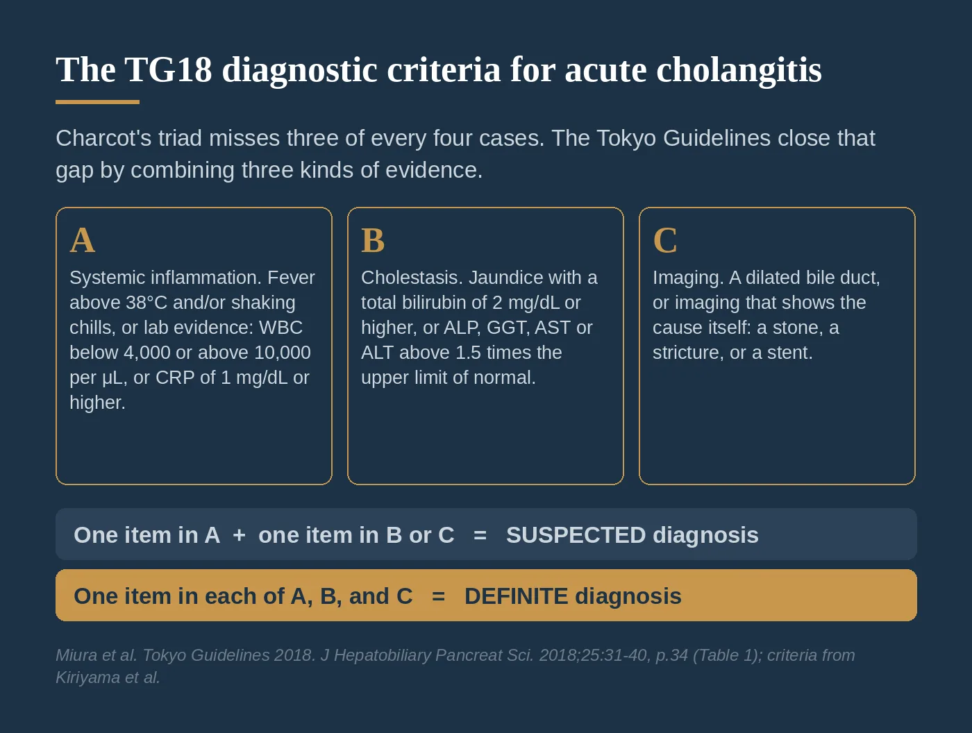
The criteria combine three kinds of evidence⁴ (p.34, Table 1):
- A, systemic inflammation: fever above 38°C and/or shaking chills, or blood work showing an inflammatory response: a white blood cell count below 4,000 or above 10,000 per μL, or a C-reactive protein of 1 mg/dL or higher
- B, cholestasis (blocked bile flow): jaundice, defined as a total bilirubin of 2 mg/dL or higher, or liver tests (ALP, GGT, AST, ALT) above 1.5 times the upper limit of normal
- C, imaging: a dilated bile duct, or imaging that shows the cause itself, such as a stone, a stricture, or a stent
One item from A plus one item from either B or C makes a suspected diagnosis. One item from each of A, B, and C makes the diagnosis definite.⁴ (p.34, Table 1)
The word definite deserves a closer look, because it is a label, not a guarantee. The two numbers above measure different things. Sensitivity of about 100% is the catch rate: of the patients who truly have cholangitis, the criteria flag essentially all of them, so a patient who does not meet the criteria is very unlikely to have the disease. Specificity of 87.4% is the clear rate: of the patients who do not have cholangitis, about 87 out of 100 are correctly cleared, which leaves roughly 13 out of 100 who meet the criteria anyway.³ In other words, the criteria are built to miss as few real cases as possible, and the cost of that design is a small group of patients who get labeled and treated for a disease they turn out not to have. So a definite diagnosis means every category was met. It does not mean the diagnosis is certain, and it does not replace the reassessment that follows.
Grading Severity: The Grade Starts the Timer
TG18 then grades every cholangitis into three tiers. Per TG18, the grade tells the team what to do and how fast to do it.⁴ (p.35, Table 3; p.36-37)
- Grade I (mild): cholangitis that meets neither of the definitions below at initial diagnosis. Initial treatment, intravenous fluids and antibiotics, is sufficient for most of these patients, and most do not need their duct drained. If the patient does not respond to initial treatment within 24 hours, the duct should be drained after all.⁴ (p.36, p.37, Table 5)
- Grade II (moderate): any two of these five: white blood cell count above 12,000 or below 4,000 per mm³; fever of 39°C or higher; age 75 or older; total bilirubin of 5 mg/dL or higher; or a low albumin (below 0.7 times the lower limit of normal).⁴ (p.35, Table 3) No organ has failed yet, but the infection is gaining ground. TG18 calls for early endoscopic or percutaneous drainage.⁴ (p.37)
- Grade III (severe): cholangitis with dysfunction of at least one organ system: cardiovascular (low blood pressure requiring pressors), neurological (confusion), respiratory (P/F ratio under 300), renal (low urine output or creatinine above 2.0 mg/dL), hepatic (INR above 1.5), or hematologic (platelets under 100,000/mm³).⁴ (p.35, Table 3) The patient can deteriorate rapidly, so TG18 calls for urgent drainage: stabilize with breathing and blood pressure support, then drain the duct as soon as possible.⁴ (p.37)
In every grade, the underlying cause still has to be treated. For a duct stone that means a sphincterotomy and stone removal, which in mild and moderate disease can often be done during the same procedure as the drainage; otherwise it follows once the patient's condition improves.⁴ (p.36-37)
One note on those pathways before we go further. They are taken directly from TG18, which lists endoscopic and percutaneous drainage side by side as options at the moderate and severe grades.⁴ (p.37) In practice the two are not used anywhere near equally in cholangitis, and I have not used percutaneous drainage as the answer for a cholangitis patient. I come back to that below, in the section on how the duct actually gets drained.
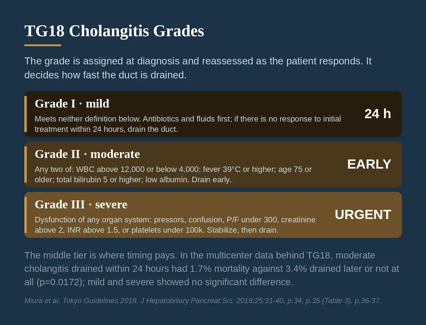
In the multicenter validation data behind TG18, mortality for moderate (Grade II) patients drained within 24 hours was 1.7%, against 3.4% for those drained later or not at all (p=0.0172). Mild and severe patients showed no significant mortality difference by drainage timing.⁴ (p.34)
Those figures compare timing inside one grade. The mortality attached to the grades themselves comes from the international study that validated these criteria in their TG13 form, which TG18 carries forward without modification.⁴ (p.34-35) Across 6,063 patients with acute cholangitis in Japan and Taiwan, 30-day mortality climbed with the grade: 1.2% for Grade I, 2.6% for Grade II, and 5.1% for Grade III.⁵ In that same dataset, Grade II patients who received urgent or early drainage died at 2.0%, against 3.7% for the Grade II patients who did not.⁵ A Grade II patient can look like they are stabilizing on antibiotics, and it is tempting to wait. The data say that is exactly the patient who benefits from being drained sooner rather than later.
Draining the Duct: What the Endoscopy Guidelines Say
Cholangitis does not wait. Treatment starts right away with intravenous fluids and antibiotics chosen to reach high levels in bile,³ but antibiotics alone do not fix a blocked pipe. The infected duct has to be drained.
There are two ways to drain it, and they are not interchangeable:
- Internal drainage (ERCP). The scope reaches the blockage from inside. The stone comes out, or a stent is placed across the blockage, and bile goes back to draining the way it always has, into the intestine. Nothing exits the body.
- External drainage (percutaneous transhepatic drainage). A needle, and then a catheter, is passed through the skin and through the liver into a bile duct above the blockage. The infected bile drains out of the body into a collection bag until the blockage can be fixed.
The 2021 ASGE guideline on managing cholangitis is direct about how that drainage should happen:
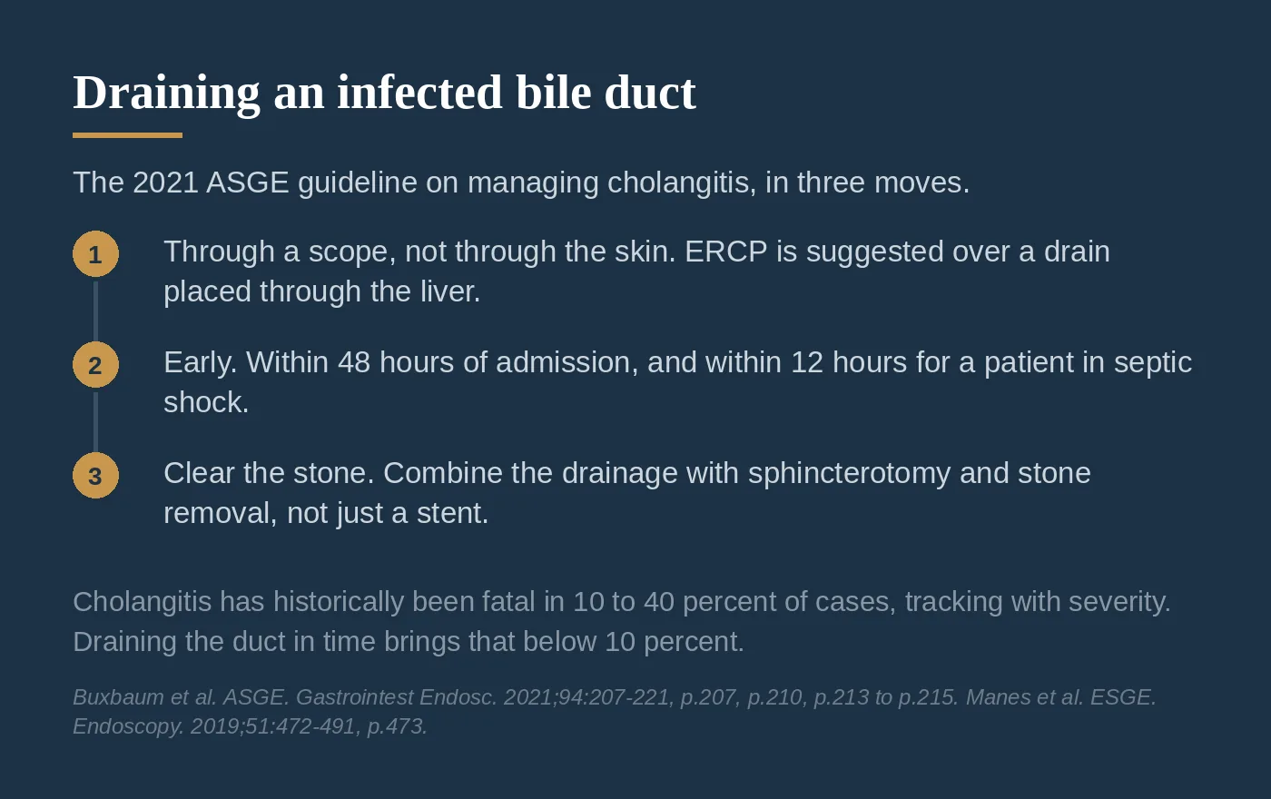
- Drain internally, through the scope, not externally through the skin. The guideline suggests ERCP over percutaneous transhepatic drainage. Patients drained by ERCP left the hospital sooner (a mean stay of 11.7 days against 23.1) and had fewer adverse events (5% against 10%, a difference driven mostly by bleeding).⁶ (p.210-211)
- Drain early. It suggests performing ERCP within 48 hours of admission rather than later. Draining within that window is associated with lower in-hospital mortality and a hospital stay shorter by several days.⁶ (p.213-214) For a patient in septic shock who is not responding to fluids, drainage within 24 hours should be considered.⁶ (p.214)
- Clear the stone, do not just relieve the pressure. When the patient is stable enough, the guideline suggests combining the drainage with a sphincterotomy and stone removal rather than placing a stent and stopping there, so the blockage is actually resolved.⁶ (p.215)
A practice note on that first recommendation. For cholangitis, external drainage is the exception. ERCP successfully decompresses the duct in about 97% of cases, and the percutaneous route is reserved for the situations where ERCP fails or cannot reasonably be done: anatomy altered by prior surgery, such as a gastric bypass, or a patient too unstable for endoscopy.⁶ (p.213, p.221) In my own practice I cannot recall a cholangitis patient who ended up with an external drain. Acute cholecystitis, which we covered in Post 5, is the opposite story: there, a percutaneous cholecystostomy tube through the skin is an option for the patient too sick for surgery. The anatomy explains the difference. In cholecystitis the infection sits in the gallbladder, at the end of a blocked duct (the cystic duct) that no endoscope can pass, so the route through the skin is the practical way in. In cholangitis the infection is in the common bile duct, so the internal route is standard.
Done well, endoscopic drainage clears cholangitis in about 94% to 98% of cases.³ That matters because cholangitis is lethal when it is left alone: historically, mortality has run from 10% to 40% and tracks closely with severity,⁶ (p.207) but it falls below 10% once the duct is drained in time.³
Two Guidelines, Two Clocks
Read the last two sections side by side and you will notice they do not fully agree.
TG18 ties the clock to the grade. Mild disease gets antibiotics first, with drainage only if the patient fails to respond within 24 hours. Moderate disease gets early drainage. Severe disease gets urgent drainage as soon as the patient is stabilized.⁴ (p.36-37) European guidance attaches hours to those words: drainage as soon as possible for severe disease, and within 12 hours for septic shock; within 48 to 72 hours for moderate disease; electively for mild disease.⁷ (p.473) The ASGE puts one clock on everyone. ERCP within 48 hours of admission, whatever the grade. Its review found insufficient data to stratify the timing question by severity, and the benefit held in both the milder and the more severe groups it could examine.⁶ (p.213-214) Going faster than 48 hours shortened the hospital stay but did not further reduce mortality or organ failure, with one exception: the patient in septic shock who is not responding to fluids, where drainage within 24 hours should be considered.⁶ (p.213-214)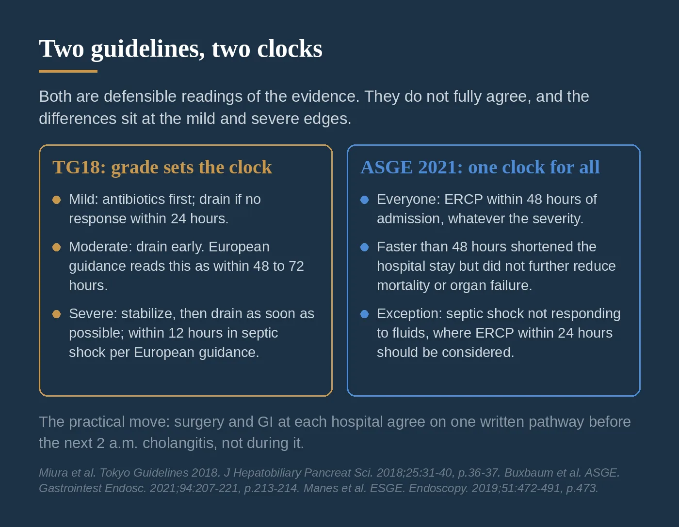
For many patients the two paths land in the same place: a moderate cholangitis drained early satisfies both documents. The differences sit at the edges. In mild disease, TG18 permits a trial of antibiotics (presumably the stone or blockage passes or resolves on its own). In severe disease, TG18's "as soon as possible" and the European 12-hour clock are faster than 48 hours, and the ASGE's own septic shock exception moves in the same direction.⁴ (p.36-37) ⁶ (p.213-214) ⁷ (p.473)
Here is my practical recommendation: the surgeons and the gastroenterologists at your hospital should determine the hospital's pathway in writing before the next 2 a.m. cholangitis, not during it. Whether your institution runs on the TG18 grade or on a flat 48-hour clock changes when the ERCP team activates and what counts as an acceptable wait. Both documents are defensible readings of the evidence. What is not defensible is two teams at the same bedside working from two different clocks and discovering it in real time. Agree on the pathway, write it down, and revisit it when the guidelines are updated.
Cholecystectomy: The Ultimate Therapy
Clearing the stone or draining the infection treats the immediate emergency, but it does not address where the stones came from. The gallbladder is the source. Clearing the duct removes the stone that is there now; it does nothing about the gallbladder that keeps forming new ones. That is why, once the acute problem is controlled, the definitive treatment is removing the gallbladder. Taking it out is what stops another stone from making the same trip and causing the same problem again.
Three practical points round this out.
First, clear the duct even if the stone is quiet. A stone in the duct should be cleared whether or not it is causing symptoms, because duct stones left in place carry a high rate of later complications, including pancreatitis, cholangitis, and obstruction.⁷ (p.474) Second, take the gallbladder out after the duct is cleared. A meta-analysis of eight studies, seven of them randomized trials, compared 12,717 patients who had a cholecystectomy after endoscopic sphincterotomy against patients whose gallbladder was left in place. Cholecystectomy cut the rate of later pancreatitis from 2.27% to 0.83% (risk ratio 0.38), cholangitis and jaundice by a similar margin (risk ratio 0.31), and cholecystitis or biliary colic by roughly three quarters (risk ratio 0.28). Mortality did not differ significantly between the groups (risk ratio 0.73, p=0.07). The benefit held in the high-risk patients as well.⁸ Third, do it during the same hospital stay when you can. A 2025 review of a national readmissions analysis of 124,964 admissions for gallstone-related cholangitis found that patients who had the gallbladder removed during the index admission were readmitted within 30 days about half as often as those sent home for an interval operation (5.56% against 11.50%), and died less often during that admission (1.18% against 3.17%). The same-admission operation added about a day of length of stay and cost more up front, and the interval group gave that back in readmissions. Even so, fewer than 15% of eligible patients get the same-admission operation.⁹For more on gallbladder disease and when removal is recommended, see my gallbladder patient guide. Missed the earlier posts in this series? You can find them all on the blog.
Up next: gallstone pancreatitis, what happens when a stone blocks more than just the bile duct.
References
1. McNicoll CF, Pastorino A, Farooq U, Froehlich MJ, St Hill CR. Choledocholithiasis. StatPearls [Internet]. Treasure Island, FL: StatPearls Publishing; updated July 10, 2023. https://www.ncbi.nlm.nih.gov/books/NBK441961/
2. ASGE Standards of Practice Committee; Buxbaum JL, Abbas Fehmi SM, Sultan S, et al. ASGE guideline on the role of endoscopy in the evaluation and management of choledocholithiasis. Gastrointest Endosc. 2019;89(6):1075-1105.e15. https://doi.org/10.1016/j.gie.2018.10.001
3. Virgile J, Marathi R. Cholangitis. StatPearls [Internet]. Treasure Island, FL: StatPearls Publishing; updated July 3, 2023. https://www.ncbi.nlm.nih.gov/books/NBK558946/
4. Miura F, Okamoto K, Takada T, et al. Tokyo Guidelines 2018: initial management of acute biliary infection and flowchart for acute cholangitis. J Hepatobiliary Pancreat Sci. 2018;25(1):31-40. https://doi.org/10.1002/jhbp.509
5. Kiriyama S, Takada T, Hwang TL, et al. Clinical application and verification of the TG13 diagnostic and severity grading criteria for acute cholangitis: an international multicenter observational study. J Hepatobiliary Pancreat Sci. 2017;24(6):329-337. https://doi.org/10.1002/jhbp.458
6. Buxbaum JL, Buitrago C, Lee A, et al. ASGE guideline on the management of cholangitis. Gastrointest Endosc. 2021;94(2):207-221.e14. https://doi.org/10.1016/j.gie.2020.12.032
7. Manes G, Paspatis G, Aabakken L, et al. Endoscopic management of common bile duct stones: European Society of Gastrointestinal Endoscopy (ESGE) guideline. Endoscopy. 2019;51(5):472-491. https://doi.org/10.1055/a-0862-0346
8. Xu J, Yang C. Cholecystectomy outcomes after endoscopic sphincterotomy in patients with choledocholithiasis: a meta-analysis. BMC Gastroenterol. 2020;20(1):229. https://doi.org/10.1186/s12876-020-01376-y
9. Paramythiotis D, Tsavdaris D, Karlafti E. Reassessing cholecystectomy timing in gallstone-related acute cholangitis. World J Gastrointest Endosc. 2025;17(5):106473. https://doi.org/10.4253/wjge.v17.i5.106473
Page numbers cited in the text, such as "p.34", refer to the printed page numbers in the published articles: J Hepatobiliary Pancreat Sci volume 25 for reference 4 (the TG18 cholangitis diagnostic criteria and severity grading tables are reproduced there, labeled TG18/TG13), Gastrointestinal Endoscopy volume 94 for reference 6, and Endoscopy volume 51 for reference 7.