In the last post, we covered biliary colic: the pain that comes and goes when a stone briefly blocks the gallbladder's outlet during a squeeze, then either falls back into the gallbladder or passes through the bile system into the duodenum. So what happens when the stone does neither?
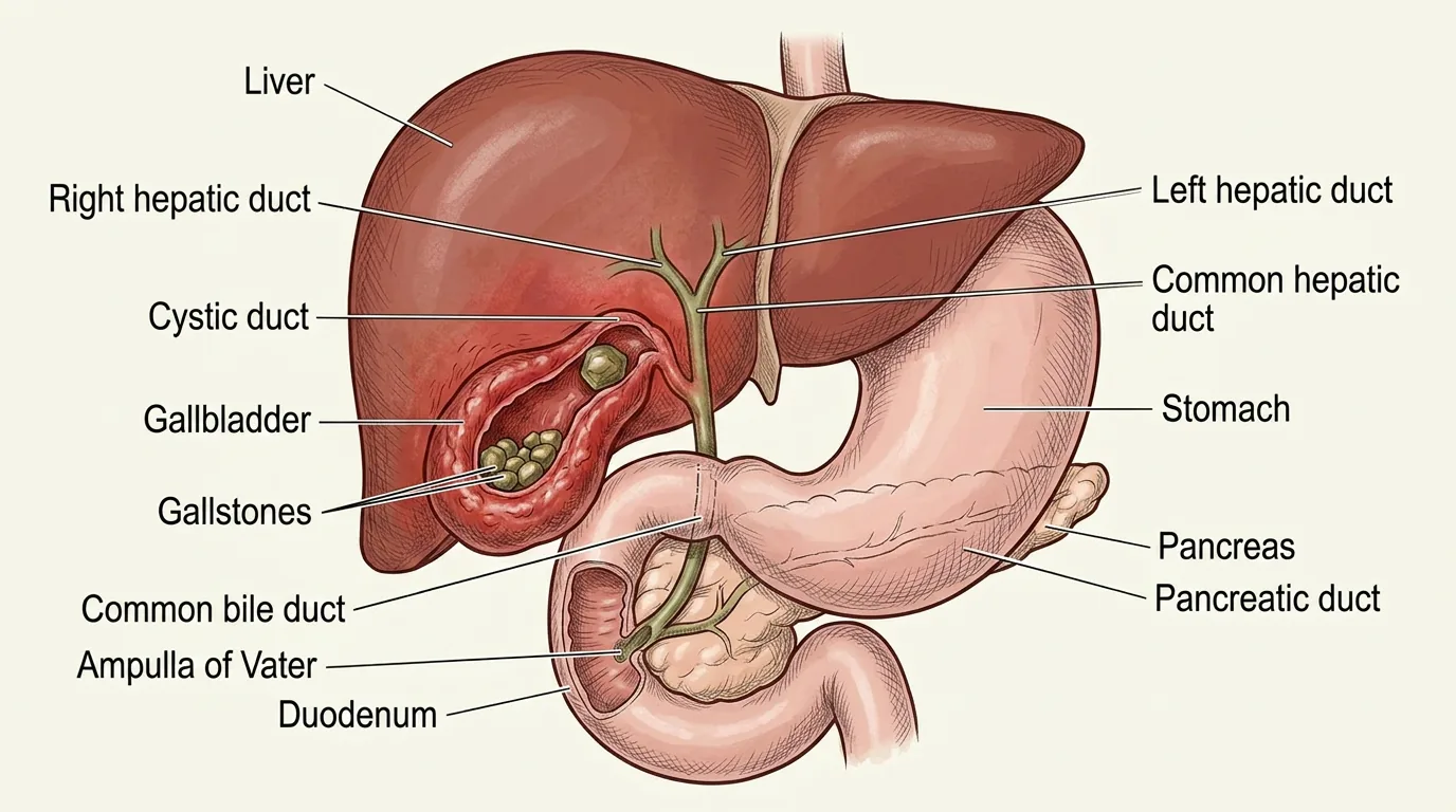
From Blockage to Inflammation
The gallbladder has one narrow opening at its neck, leading into the cystic duct. That single tube is both the way in and the way out. When a stone wedges there and stays, the gallbladder is sealed.
What follows is a chain reaction:
- The lining keeps producing fluid into a space that has nowhere to drain
- The bile already inside becomes concentrated and chemically irritating
- Pressure climbs inside the gallbladder until it begins choking off the blood supply to the wall itself
- Tissue that is stagnant and starved of blood is easy for bacteria to colonize, and infection often follows¹
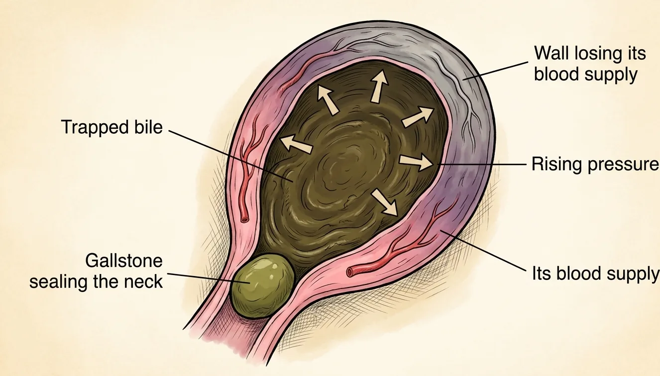
That is acute cholecystitis: an inflamed, frequently infected gallbladder that cannot empty.
How Cholecystitis Differs From Biliary Colic
The two can feel similar at the start, which is part of why patients often aren't sure how serious it is. A few things separate them:
- Biliary colic peaks within about 15 to 60 minutes and then eases off as the stone falls back into the gallbladder or passes through the bile system into the duodenum
- Acute cholecystitis causes pain that lasts more than six hours and does not resolve on its own²
- Fever and vomiting are common with cholecystitis but not typical with simple colic²
- On exam, pressing under the right ribs while a patient takes a deep breath produces pain sharp enough to catch the breath. This is Murphy's sign, which the Tokyo Guidelines describe as specific to acute cholecystitis³ (p.33)
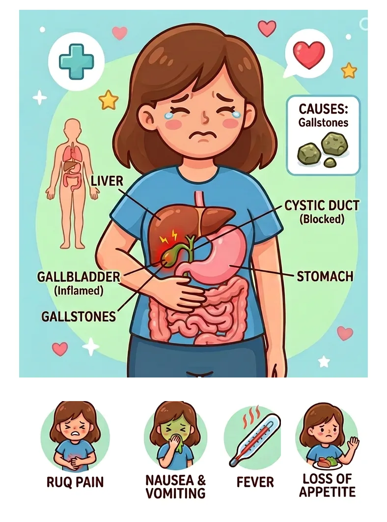
The practical takeaway for patients: colic that used to last an hour or two and now hasn't let up, especially with a fever, is a reason to be seen the same day.
One more thing worth saying, because patients worry about it. We treat your pain early. There was a long-standing concern that pain medicine would mask the physical findings and hide the diagnosis. A randomized trial of intravenous morphine against placebo in emergency-room patients with abdominal pain found no difference in how often the diagnosis was made correctly, and the Tokyo Guidelines now recommend that analgesia be given proactively and early³ (p.33). You do not have to prove how much it hurts by going without relief.
What the Tokyo Guidelines Are, and Why We Follow Them
I cite the Tokyo Guidelines throughout this post, so it is worth explaining what they are.
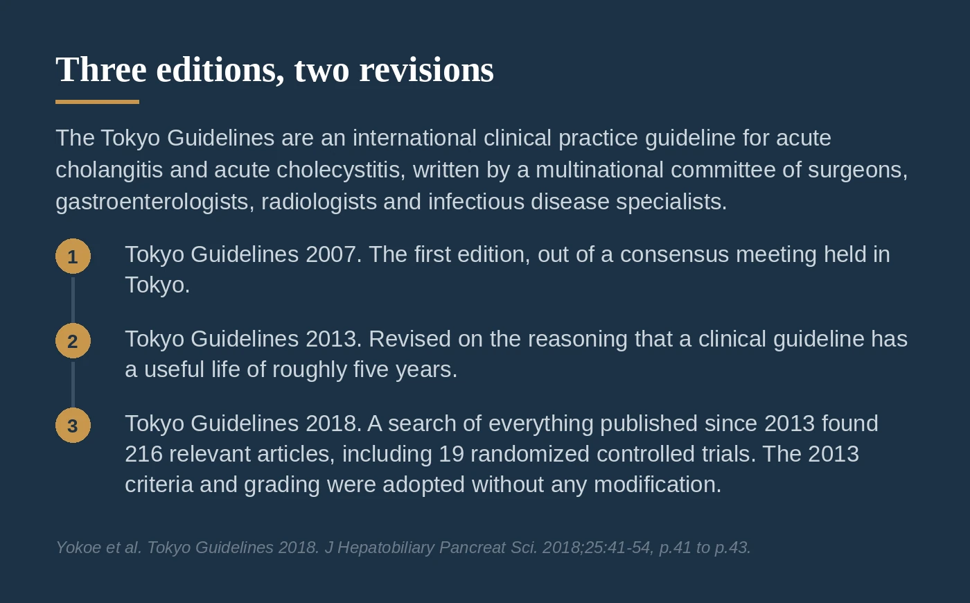
They are an international clinical practice guideline for acute cholangitis and acute cholecystitis, produced by a committee of surgeons, gastroenterologists, radiologists, and infectious disease specialists from Japan, Taiwan, Korea, the United States, and Europe. The first edition came out of a consensus meeting held in Tokyo and was published as the Tokyo Guidelines 2007⁴ (p.42).
They have been revised twice since, which gives three editions in total. The committee revised TG07 in 2013 on the reasoning that a clinical guideline has a useful lifespan of roughly five years⁴ (p.42). For the 2018 revision, they searched everything published since 2013 and identified 216 relevant articles, including 19 randomized controlled trials⁴ (p.42).
Two things make them the reference standard for this disease.
The first is that nothing else fills the role. The TG18 authors put it directly: to date, no diagnostic criteria for acute cholecystitis meriting that title have been established other than the Tokyo Guidelines⁴ (p.43). Other groups have published guidance on managing gallbladder disease, but they have not proposed diagnostic criteria or severity grading of their own⁴ (p.43, p.48).
The second is the size of the validation behind them. A Japan and Taiwan collaborative project gathered data on more than 7,000 cases of biliary infection, including over 5,000 patients with acute cholecystitis, specifically to test whether the TG13 criteria and grading held up in practice⁴ (p.42). They did. TG18 reviewed that evidence and adopted the TG13 diagnostic criteria and severity grading without any modification⁴ (p.41, p.43). A guideline that survives a 5,000-patient test unchanged is a guideline worth using.
How We Diagnose It
The Tokyo Guidelines do not leave the diagnosis to impression. They set it out in three parts⁴ (p.43):
- A, local signs of inflammation: Murphy's sign, or a right upper quadrant mass, pain, or tenderness
- B, systemic signs of inflammation: fever, elevated C-reactive protein, or elevated white blood cell count
- C, imaging findings characteristic of acute cholecystitis
One item from A plus one item from B makes a suspected diagnosis. Add C and the diagnosis is definite. Validated against a series of 451 patients, those criteria performed at 91.2% sensitivity and 96.9% specificity⁴ (p.42, p.43).
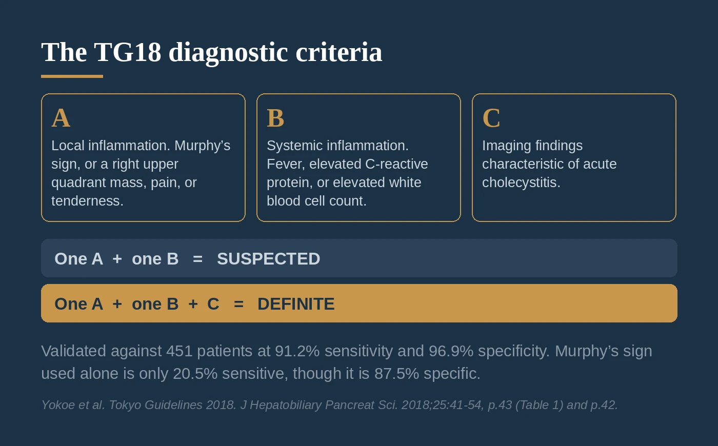
That structure matters because no single finding carries the diagnosis on its own. Murphy's sign, the exam finding everyone learns first, is only 20.5% sensitive when used alone, though it is 87.5% specific⁴ (p.42). A positive Murphy's sign is useful evidence. Its absence does not rule out cholecystitis.
Ultrasound is the first test. The Tokyo Guidelines recommend it as the first-choice imaging method, not because it is the most accurate test available, but because it is inexpensive, non-invasive, and available everywhere⁴ (p.43). Pooled across studies it runs about 81% sensitive (95% CI 0.75 to 0.87) and 83% specific (95% CI 0.74 to 0.89)⁴ (p.43).
What we are looking for on that scan⁴ (p.45):
- Gallbladder wall thickening of 4 mm or more
- An enlarged gallbladder, long axis 8 cm or more, short axis 4 cm or more
- Gallstones or retained debris
- Fluid collecting around the gallbladder
- Pain directly under the probe as it presses over the gallbladder, the sonographic Murphy's sign³ (p.33)
When ultrasound does not answer the question, what comes next depends on what you are still asking.
The Tokyo Guidelines name MRI with MRCP as the next study. It runs about 85% sensitive and 81% specific, and MRCP can show biliary anatomy without contrast⁴ (p.45). TG18 adds its own caveat: those pooled numbers come from four studies done around the turn of the millennium, before contrast-enhanced MRI and MRCP were in use⁴ (p.45).
In practice we go from ultrasound to a HIDA scan, not to MRI. If the question is still whether this is cholecystitis, the question is functional: is the cystic duct blocked? Ultrasound and MRCP both answer anatomic questions, HIDA can specifically look at cystic duct blockage, and, for diagnosing cholecystitis, it is a more sensitive test than MRCP.
MRCP earns its place when the question really is anatomic, such as a suspected stone in the common bile duct. Contrast-enhanced CT or MRI is the right call when gangrenous cholecystitis is the concern, because wall irregularity and rupture are routinely underestimated on ultrasound⁴ (p.49).
Where the HIDA Scan Earns Its Place
If ultrasound is only 81% sensitive, the obvious question is why we don't lead with something better. There is something better, and it is worth knowing when to reach for it.
A HIDA scan, formally hepatobiliary scintigraphy, is a nuclear medicine study. A small amount of tracer is injected, the liver pulls it out of the blood and excretes it into bile, and a camera watches where the bile goes over the next hour or so. If the tracer fills the gallbladder, the cystic duct is open. If the gallbladder never fills, it is blocked, which is the defining event in acute cholecystitis.
That is a different question from the one ultrasound answers. Ultrasound looks at structure: how thick is the wall, is there fluid around it, are there stones. HIDA looks at function: does this gallbladder still connect to the biliary tree?
In the meta-analysis TG18 reproduces, pooling 57 studies and 5,859 patients, cholescintigraphy was 96% sensitive (95% CI 94 to 97) and 90% specific (95% CI 86 to 93). Its sensitivity was significantly higher than ultrasound at 81% and MRI at 85%, while the differences in specificity were not significant⁵. TG18 says the same thing in its own words: every study comparing HIDA with ultrasound has found the diagnostic yield higher for HIDA⁴ (p.43).
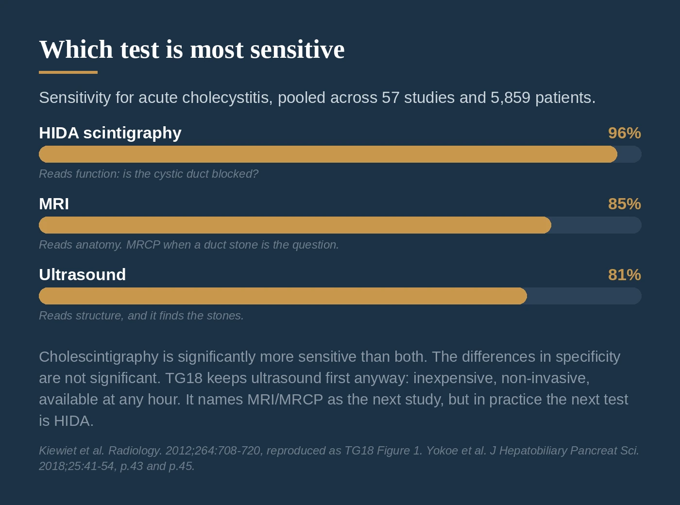
So why is ultrasound still first? Because HIDA's advantage is sensitivity, not specificity, and sensitivity is what you need when you are trying to rule out rather than rule in. Ultrasound is cheap, non-invasive, available at any hour, and it finds the stones, which HIDA cannot. TG18 recommends it first for those practical reasons and says so directly, acknowledging its limited diagnostic yield in the same sentence⁴ (p.43).
HIDA earns its place in a specific situation: the ultrasound is equivocal, and the clinical picture still fits. A negative HIDA in that setting is a genuine answer, in a way that a negative ultrasound is not. That is why it, and not MRI, is the test we actually order next.
We will cover the full imaging workup in its own post later in this series.
Severity Grading, and Why It Matters
The Tokyo Guidelines grade acute cholecystitis into three levels, and the grade changes the plan⁴ (p.49):
- Grade I (mild): a healthy patient, no organ dysfunction, mild inflammatory changes. Cholecystectomy is a safe, low-risk operation.
- Grade II (moderate): any one of these. White count above 18,000/mm³. A palpable tender right upper quadrant mass. Symptoms lasting more than 72 hours. Or marked local inflammation, which includes gangrenous cholecystitis, pericholecystic abscess, hepatic abscess, biliary peritonitis, or emphysematous cholecystitis.
- Grade III (severe): dysfunction of any one organ system. Cardiovascular (needing pressors), neurological, respiratory (P/F ratio under 300), renal (creatinine above 2.0 mg/dL), hepatic (INR above 1.5), or hematologic (platelets under 100,000/mm³).
Here is what the outcomes look like. In a cohort of 3,721 patients, 30-day mortality was 1.1% for Grade I (15 deaths of 1,339), 0.8% for Grade II (13 of 1,702), and 5.4% for Grade III (37 of 680), with an overall p-value below 0.001⁴ (p.47).
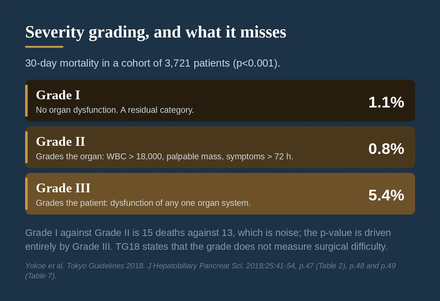
Read that carefully, because Grade I and Grade II look backwards. They are not. Two things are going on.
First, the Grade I and Grade II figures are 15 deaths against 13 deaths. That difference is noise, and the p-value below 0.001 is driven entirely by Grade III, where the rate is roughly five times either of the others.
Second, and more useful clinically: Grade II is defined by how inflamed the gallbladder is, not by how sick the patient is. A high white count, a palpable mass, symptoms past 72 hours, an abscess. Those describe the organ. A patient can have a furiously inflamed gallbladder and an otherwise excellent physiologic reserve. Grade I, meanwhile, is a residual category: it collects everyone who meets neither the Grade II nor the Grade III criteria, and that includes frail and elderly patients whose gallbladder inflammation is unremarkable but whose ability to survive an illness is not.
Only Grade III measures the patient rather than the organ, because organ dysfunction is its definition. That is why Grade III separates so sharply from the other two, and why the grading system predicts mortality well at the top and poorly in the middle.
The grade tracks other outcomes more consistently. Conversion to open surgery in one 445-patient series ran 7.0%, 25.6%, and 34.6% across the three grades (p=0.001), and complications ran 2.9%, 3.1%, and 11.1% (p=0.003)⁴ (p.47, p.48). One thing the grade does not measure is how technically difficult the operation will be. TG18 states that directly⁴ (p.48).
Why This One Needs Surgery
Unlike gallstones that never cause symptoms, acute cholecystitis is not something we watch and wait on. Once the diagnosis is confirmed, treatment is antibiotics and removal of the gallbladder, ideally during the same hospital admission.
The Tokyo Guidelines are direct about it: if a patient is able to withstand surgery, they propose early surgery regardless of how long symptoms have been going on⁶ (p.59). That last part is a change worth noticing. The old teaching was a hard 72-hour window, after which you cooled the patient down with antibiotics and brought them back in six weeks. TG18 says that for patients past 72 hours, "there still are benefits to performing surgery early"⁶ (p.63).
Their own meta-analysis of 15 randomized controlled trials found⁶ (p.60, p.61):
- Total hospital stay was shorter with early surgery (p<0.0001)
- Overall cost was lower (p=0.002)
- Operating time was not significantly different (p=0.16 and p=0.06)
- Bile duct injury showed no significant difference (p=0.45 and p=0.72)
That last line needs a caveat, and TG18 supplies it in the same paragraph: the pooled number of patients is far too small to conclude anything about bile duct injury in either direction. Their words are "Absence of evidence is not evidence of absence"⁶ (p.61). A separate meta-analysis in the British Journal of Surgery reached the same broad conclusion in favor of early surgery⁷. So the fair statement is this: early surgery clearly shortens hospitalization and lowers cost, and there is no signal that it is more dangerous, but nobody has run a trial large enough to answer the bile duct injury question on its own.
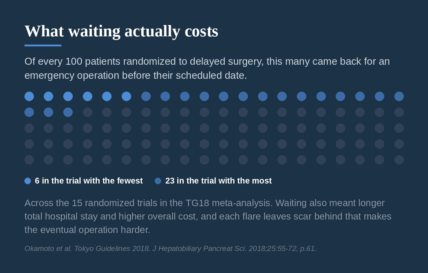
Here is the number that carries the argument for me. In those 15 trials, between 6% and 23% of the patients assigned to wait came back for an emergency operation before their scheduled date, because their symptoms flared again during the waiting period⁶ (p.61). And each flare leaves scar behind. TG18 notes that tissues become progressively more scarred with repeated episodes, which makes the eventual operation harder and riskier⁶ (p.61).
Waiting does not make the operation safer. It adds hospital time, it carries a real chance of an unplanned emergency operation, and it leaves in place a gallbladder that can progress to gangrene or, less commonly, perforation, a complication that carries a 30% mortality when it occurs¹.
When the Patient Is Too Sick for Surgery: the C-Tube
Not everyone can go to the operating room. Some patients arrive in septic shock, on a ventilator, on blood thinners that cannot be reversed quickly, or with heart or lung disease severe enough that anesthesia is the bigger threat. For them, TG18 provides a second path: drain the gallbladder instead of removing it.
The usual method is a cholecystostomy tube, which most of us shorten to C-tube. An interventional radiologist passes a thin tube through the skin and through the edge of the liver into the gallbladder, using ultrasound or CT for guidance, and leaves it in place to drain. It relieves the pressure and lets the infection out without an operation and usually without general anesthesia.
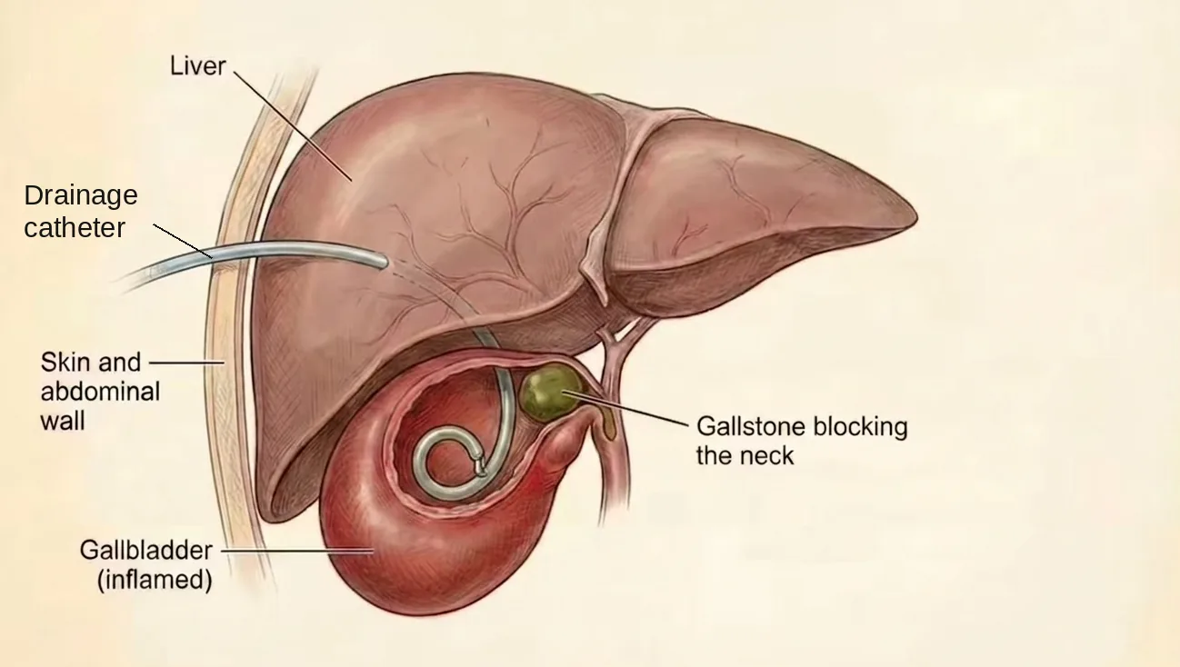
Where it fits in the TG18 flowcharts:
- Grade II, when antibiotics and supportive care fail to bring the inflammation under control and the patient is high risk. TG18 sets that bar at a Charlson comorbidity index of 6 or more, or an ASA physical status of class III or higher. Both conditions have to be met before the flowchart sends you to urgent or early gallbladder drainage⁶ (p.66, Figure 9).
- Grade III, when the patient does not clear the bar for early cholecystectomy even with intensive care. Urgent or early gallbladder drainage should be performed, with elective cholecystectomy considered later, once the acute illness has improved⁶ (p.67).
A C-tube is a bridge, not a cure. The stones are still there and the gallbladder is still diseased, so most patients come back for a cholecystectomy once they have recovered. When they come back is still an open question: TG18 reviewed the evidence on timing after a C-tube and found no quality data and no consensus⁶ (p.63). Two studies that compared operating after a 4 to 6 week wait against operating early after the tube reported better bleeding, operative times, and conversion rates with the longer wait⁶ (p.63). In practice the treating physician judges the timing against that individual patient's risk.
A note on antibiotics. For Grade I and Grade II disease, TG18 recommends antibiotics only before and at the time of surgery. They stop when the gallbladder comes out⁸ (p.11). Two randomized trials of postoperative antibiotics against none in mild-to-moderate disease found a risk difference for postoperative infection of 0.01 (95% CI −0.04 to 0.06)⁸ (p.11). Grade III is different: 4 to 7 days after the source of infection is controlled⁸ (p.11). TG18 is candid that this figure rests on expert opinion rather than strong data, and records a dissent at the consensus meeting saying as much⁸ (p.11). If blood cultures grow gram-positive organisms such as Enterococcus or Streptococcus, the recommendation extends to a minimum of two weeks, because of the risk of endocarditis⁸ (p.10).
We will walk through exactly how that operation is performed later in this series, in Part 4: Surgery.
For more on gallbladder disease and when removal is recommended, see my gallbladder patient guide. Missed the earlier posts in this series? You can find them all on the blog. If you want the surgical technique side of this, I wrote about the critical view of safety separately.
Up next: choledocholithiasis, what happens when a stone escapes the gallbladder and gets into the bile duct itself.
References
1. Jones MW, Santos G, Patel PJ, O'Rourke MC. Acute Cholecystitis. StatPearls [Internet]. Treasure Island, FL: StatPearls Publishing; updated July 6, 2025. https://www.ncbi.nlm.nih.gov/books/NBK459171/
2. Acute Cholecystitis. Merck Manual Professional Edition. Modified October 2025. https://www.merckmanuals.com/professional/hepatic-and-biliary-disorders/gallbladder-and-bile-duct-disorders/acute-cholecystitis
3. Miura F, Okamoto K, Takada T, et al. Tokyo Guidelines 2018: initial management of acute biliary infection and flowchart for acute cholangitis. J Hepatobiliary Pancreat Sci. 2018;25(1):31-40. https://doi.org/10.1002/jhbp.509
4. Yokoe M, Hata J, Takada T, et al. Tokyo Guidelines 2018: diagnostic criteria and severity grading of acute cholecystitis (with videos). J Hepatobiliary Pancreat Sci. 2018;25(1):41-54. https://doi.org/10.1002/jhbp.515
5. Kiewiet JJS, Leeuwenburgh MMN, Bipat S, Bossuyt PMM, Stoker J, Boermeester MA. A systematic review and meta-analysis of diagnostic performance of imaging in acute cholecystitis. Radiology. 2012;264(3):708-720. https://doi.org/10.1148/radiol.12111561
6. Okamoto K, Suzuki K, Takada T, et al. Tokyo Guidelines 2018: flowchart for the management of acute cholecystitis. J Hepatobiliary Pancreat Sci. 2018;25(1):55-72. https://doi.org/10.1002/jhbp.516
7. Wu XD, Tian X, Liu MM, Wu L, Zhao S, Zhao L. Meta-analysis comparing early versus delayed laparoscopic cholecystectomy for acute cholecystitis. Br J Surg. 2015;102(11):1302-1313. https://pubmed.ncbi.nlm.nih.gov/26265548/
8. Gomi H, Solomkin JS, Schlossberg D, et al. Tokyo Guidelines 2018: antimicrobial therapy for acute cholangitis and cholecystitis. J Hepatobiliary Pancreat Sci. 2018;25(1):3-16. https://doi.org/10.1002/jhbp.518
9. Wakabayashi G, Iwashita Y, Hibi T, et al. Tokyo Guidelines 2018: surgical management of acute cholecystitis: safe steps in laparoscopic cholecystectomy for acute cholecystitis (with videos). J Hepatobiliary Pancreat Sci. 2018;25(1):73-86. https://doi.org/10.1002/jhbp.517
Page numbers cited in the text, such as "p.43", refer to the printed page numbers in J Hepatobiliary Pancreat Sci volume 25, so they can be located directly in the published article.