In the last post, we covered choledocholithiasis and cholangitis: a stone that leaves the gallbladder, lodges in the common bile duct, and blocks the drainage of the whole liver. Just before the common bile duct empties into the small intestine (duodenum), it joins the duct that drains the pancreas. If a stone stops at that junction both ducts become blocked. If pancreatitis inflammation ensues, this is termed gallstone (biliary) pancreatitis.
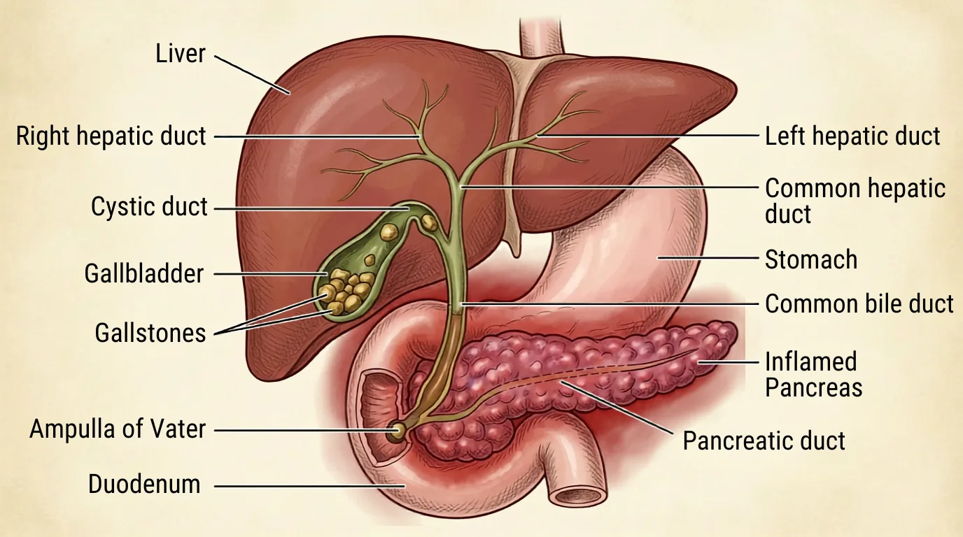
Gallstones are the most common cause of acute pancreatitis in the United States, and alcohol is second. StatPearls puts the gallstone share at roughly 35% to 40% of cases.¹ The American College of Gastroenterology guideline puts it higher, at 40% to 70%.²
What Actually Happens to the Pancreas
The pancreas makes digestive enzymes. Those enzymes stay inactive until they reach the intestine, where they are switched on to break down food.
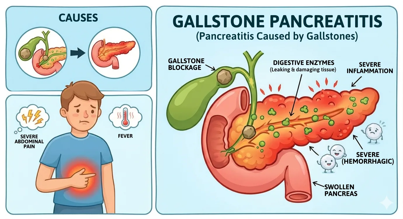
A stone lodged at the ampulla of Vater breaks that sequence. The stone transiently obstructs the ampulla, bile refluxes alongside pancreatic secretions into the pancreatic duct, and the digestive enzymes activate prematurely, inside the pancreas itself.¹ The organ begins to digest its own tissue. This is termed autodigestion.
Small stones are the ones that do this. Stones under about 5 mm, and biliary sludge, are more likely to migrate out of the gallbladder and reach the ampulla.¹ Larger stones tend to stay in the gallbladder.
The Stone Is Usually Gone by the Time We Look
By the time the diagnosis is made, the stone that caused the attack has often already moved on. It has left the gallbladder, so it does not go back there. It either stays in the common bile duct or passes into the duodenum.
The obstruction that starts gallstone pancreatitis can be transient.¹ Once autodigestion begins, the inflammation continues on its own whether or not the stone is still there.
Small duct stones do pass without help. In one series cited by the European Society of Gastrointestinal Endoscopy guideline, 24 of 46 patients who had a filling defect on an intraoperative cholangiogram passed the stone with no intervention³ (p.474). That series was not a pancreatitis cohort, so it does not give a passage rate for this disease.
This can definitely lead to some confusion. Patients are told they have a stone problem, then told there is no stone to remove, then told they still need surgery. All three are correct.
Making the Diagnosis
The diagnosis of acute pancreatitis follows the Revised Atlanta Classification and requires at least two of these three findings:¹
- Abdominal pain consistent with the disease, typically in the upper abdomen and radiating to the back
- A serum amylase or lipase at least three times the upper limit of normal
- Characteristic findings on imaging, by ultrasound, CT, or MRI
A patient with the classic pain and a lipase five times normal has acute pancreatitis. Imaging is how we find the cause, and for gallstone pancreatitis that means an abdominal ultrasound to look at the gallbladder, plus a liver panel (AST, ALT, bilirubin).
Lipase is the better test. The ACG guideline states that serum amylase alone cannot be used reliably for the diagnosis and that lipase is preferred.² Once the diagnosis is established, there is no reason to keep following the amylase or lipase. The number has no relationship to severity, prognosis, or the decision about when to feed or discharge the patient.²
How Sick Is Sick
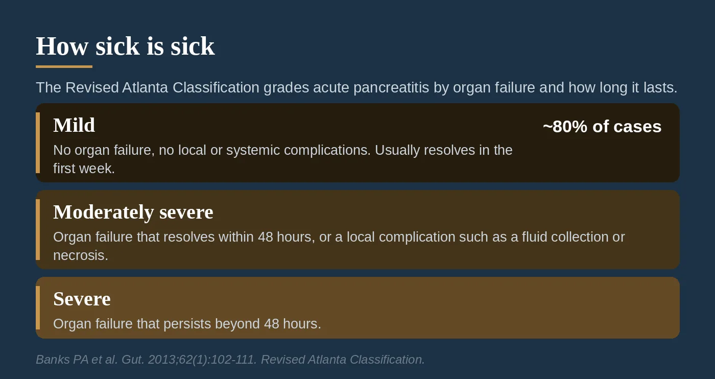
The Revised Atlanta Classification sorts acute pancreatitis into three tiers, defined by organ failure rather than by how the patient looks on arrival:¹⁴
- Mild. No organ failure, no local complications, no systemic complications. Usually resolves within the first week.
- Moderately severe. Transient organ failure that resolves within 48 hours, or a local complication such as a fluid collection or sterile necrosis, or worsening of an existing medical problem.
- Severe. Persistent organ failure, meaning organ failure that does not resolve within 48 hours (single or multiple organ system).
That 48-hour mark is what separates moderately severe from severe. The same patient with the same creatinine on day one falls into a different category depending on where that creatinine sits on day three.
Approximately 80% of patients present with mild disease and are discharged within several days.¹ Moderately severe and severe disease together account for roughly 15% to 25% of cases.² Overall mortality for acute pancreatitis runs about 1% to 2%, and it separates sharply by severity: roughly 3% in mild interstitial pancreatitis and around 20% once there is necrosis.¹
What Treatment Actually Involves
There is no drug that stops pancreatic autodigestion. Treatment is supportive: fluids, pain control, and feeding the patient. Two of those three have changed in the last several years.
Fluids: moderate beats aggressive. The teaching used to be to resuscitate hard. The WATERFALL trial tested that, randomizing patients with acute pancreatitis to aggressive or moderate fluid resuscitation with lactated Ringer's.⁵ The trial was stopped early at its first interim analysis, with 249 patients enrolled. Aggressive resuscitation did not reduce progression to moderately severe or severe pancreatitis: 22.1% versus 17.3%, adjusted relative risk 1.30 (95% CI 0.78 to 2.18), p=0.32. It did cause fluid overload, in 20.5% of the aggressive group versus 6.3% of the moderate group, adjusted relative risk 2.85 (95% CI 1.36 to 5.94), p=0.004.⁵
The ACG guideline reflects this. It suggests moderately aggressive fluid resuscitation (Recommendation 3) and lactated Ringer's solution over normal saline (Recommendation 4), with an initial rate of no more than 1.5 mL/kg per hour in a patient who is not hypovolemic, and a 10 mL/kg bolus for a patient who is.² Close observation is required when administering IV fluids to prevent volume overload.
Feeding: earlier and more normal. The old approach was nothing by mouth until the pain resolved, then clear liquids, then advancing slowly. The guideline now suggests early oral feeding within 24 to 48 hours as tolerated in mild acute pancreatitis (Recommendation 10), and suggests starting with a low-fat solid diet rather than the stepwise liquid-to-solid progression (Recommendation 11).² Hunger is a reasonable indication to start. The same data holds for moderately severe pancreatitis.
In severe pancreatitis, the data is less clear. On timing, pushing feeding earlier than that does not appear to help. The PYTHON trial randomized 208 patients with acute pancreatitis at high risk for complications to nasoenteric tube feeding within 24 hours, or to an oral diet started at 72 hours with a tube only if the oral diet was not tolerated. Major infection or death occurred in 30% of the early group and 27% of the on-demand group, relative risk 1.07 (95% CI 0.79 to 1.44), p=0.76. In the on-demand group, 69% tolerated an oral diet and never needed a tube at all.⁶ When a tube is needed, the ACG guideline prefers the nasogastric route over the nasojejunal route, because safety and efficacy are comparable (Key concept 20).² Gastric access is also easier to obtain and to keep. Hunger, if the patient is awake and interactive, should guide oral intake.
Antibiotics: not prophylactically. The guideline suggests against prophylactic antibiotics in severe acute pancreatitis (Recommendation 8).² Antibiotics have a clear role in infected necrosis. They do not prevent it.
Does Everyone Need an ERCP?
No.
The reasoning goes: a stone caused this, so we should go get the stone. Sometimes the stone has already passed, and ERCP carries its own risk of causing pancreatitis.
The APEC trial tested urgent ERCP in the population where it seemed most likely to help: 232 patients with predicted severe gallstone pancreatitis, without cholangitis, randomized to urgent ERCP with sphincterotomy within 24 hours or to conservative treatment.⁷ The composite of death or major complications occurred in 38% of the urgent ERCP group and 44% of the conservative group, relative risk 0.87 (95% CI 0.64 to 1.18), p=0.37. That difference is not statistically significant, and the confidence interval is wide enough to include a meaningful benefit or a meaningful harm.
One component did differ. Cholangitis developed in 2% of the urgent ERCP patients versus 10% of the conservative patients, relative risk 0.18 (95% CI 0.04 to 0.78), p=0.010.⁷ Opening the sphincter prevented later infection of the duct. It did not change death or major complications overall.
So the rule is narrow and specific. The ACG guideline suggests medical therapy over early ERCP in acute biliary pancreatitis without cholangitis (Recommendation 5).² When pancreatitis is complicated by cholangitis, early ERCP within the first 24 hours reduces morbidity and mortality (Key concept 13).² When a duct stone is suspected but there is no cholangitis and no jaundice, the guideline directs us to screen with MRCP or endoscopic ultrasound first, and to avoid a purely diagnostic ERCP (Key concept 14).²
An infected duct, or a duct that is still obstructed, gets an ERCP. Pancreatitis by itself does not.
The Gallbladder Still Has to Come Out
Clearing the duct, or watching a stone pass on its own, treats the episode. It does nothing about the organ that made the stone. The gallbladder that produced one migrating stone will produce another.
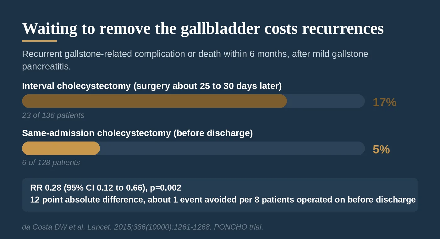
The PONCHO trial randomized 266 patients recovering from mild gallstone pancreatitis to cholecystectomy during the same hospital admission or to discharge with surgery about 25 to 30 days later.⁸ Within six months, a recurrent gallstone-related complication or death occurred in 17% of the interval group (23 of 136) and 5% of the same-admission group (6 of 128), relative risk 0.28 (95% CI 0.12 to 0.66), p=0.002.⁸
Both numbers matter. The relative risk reduction is 72%. The absolute difference is 12 percentage points, which is roughly one recurrent gallstone complication avoided for every eight patients whose gallbladder comes out before they go home.
The safety side did not offset it. Across both arms combined there were four serious surgical adverse events, one bile duct leak and one postoperative bleed in each group, and none led to death.⁸ Operating during the index admission did not make the operation more dangerous.
The ACG guideline states it as a key concept: patients with mild acute biliary pancreatitis should undergo cholecystectomy early, preferably before discharge (Key concept 21).²
Severe disease is the exception. When pancreatitis is complicated by necrosis or by peripancreatic collections, the operation gets harder and riskier. The ESGE guideline recommends performing cholecystectomy once the collections and local complications have resolved, generally beyond 6 weeks³ (p.482). For a patient who is not fit for surgery, the gallbladder can be left in place, but that is a second choice and not an equal one. ESGE states that clearing the duct without removing the gallbladder is inferior to doing both, for mortality and for recurrent biliary events, and that the gallbladder should be left in situ only in patients who are unfit for surgery³ (p.482). Endoscopic sphincterotomy lowers the rate of later biliary events, including pancreatitis, but it does not remove the gallbladder or the risk³ (p.482).
I remove gallbladders robotically, and have done so exclusively since May 2022. For a patient recovering from mild gallstone pancreatitis, taking the gallbladder out before discharge is one of the more clearly beneficial things we do in general surgery. The evidence is a randomized trial and the effect size is large.
For more on gallbladder disease and when removal is recommended, see our full guide to gallbladder disease. Missed the earlier posts in this series? You can find them all on the blog.
Up next: gallbladder and biliary tree imaging.
References
1. Wang CF, Tariq A, Chandra S. Acute Pancreatitis. StatPearls [Internet]. Treasure Island, FL: StatPearls Publishing; updated August 2, 2025. https://www.ncbi.nlm.nih.gov/books/NBK482468/
2. Tenner S, Vege SS, Sheth SG, et al. American College of Gastroenterology Guidelines: Management of Acute Pancreatitis. Am J Gastroenterol. 2024;119(3):419-437. https://doi.org/10.14309/ajg.0000000000002645
3. Manes G, Paspatis G, Aabakken L, et al. Endoscopic management of common bile duct stones: European Society of Gastrointestinal Endoscopy (ESGE) guideline. Endoscopy. 2019;51(5):472-491. https://doi.org/10.1055/a-0862-0346
4. Banks PA, Bollen TL, Dervenis C, et al. Classification of acute pancreatitis 2012: revision of the Atlanta classification and definitions by international consensus. Gut. 2013;62(1):102-111. https://doi.org/10.1136/gutjnl-2012-302779
5. de-Madaria E, Buxbaum JL, Maisonneuve P, et al. Aggressive or Moderate Fluid Resuscitation in Acute Pancreatitis. N Engl J Med. 2022;387(11):989-1000. https://doi.org/10.1056/NEJMoa2202884
6. Bakker OJ, van Brunschot S, van Santvoort HC, et al. Early versus on-demand nasoenteric tube feeding in acute pancreatitis (PYTHON). N Engl J Med. 2014;371(21):1983-1993. https://doi.org/10.1056/NEJMoa1404393
7. Schepers NJ, Hallensleben NDL, Besselink MG, et al. Urgent endoscopic retrograde cholangiopancreatography with sphincterotomy versus conservative treatment in predicted severe acute gallstone pancreatitis (APEC): a multicentre randomised controlled trial. Lancet. 2020;396(10245):167-176. https://doi.org/10.1016/S0140-6736(20)30539-0
8. da Costa DW, Bouwense SA, Schepers NJ, et al. Same-admission versus interval cholecystectomy for mild gallstone pancreatitis (PONCHO): a multicentre randomised controlled trial. Lancet. 2015;386(10000):1261-1268. https://doi.org/10.1016/S0140-6736(15)00274-3
Recommendation and Key concept numbers cited for reference 2 refer to the numbered statements in the published ACG guideline, which is open access at the link above. Page numbers cited for reference 3 are the printed page numbers in Endoscopy volume 51, read from the published PDF. For orientation, the summary recommendations sit on p.473 and the reference list begins on p.484.