Last week I walked through a gallbladder operation in the order I do it. Gaining access, three more incisions, the first look, the duct and the artery, off the liver and out.
That post left something out on purpose. Between step three and step four there is a pause. That pause is the most important part of the operation.
The structure that must stay untouched
Your liver makes bile all day, and all of it leaves by one channel. That channel picks up different names along the way. The right and left hepatic ducts come out of the liver and join to form the common hepatic duct, which becomes the common bile duct, which empties into your intestine and helps you digest fat. Several names, one channel, and all of it has to be intact when I am finished.
Your gallbladder is not part of that channel. It hangs off the side of it on a short branch called the cystic duct, and it stores bile between meals.
To remove the gallbladder I clip and cut the cystic duct, and I clip, cauterize and cut the cystic artery. Both of those belong to the gallbladder. Neither one carries bile from your liver to your intestine. Being certain of that before anything gets divided is what the rest of this post is about.
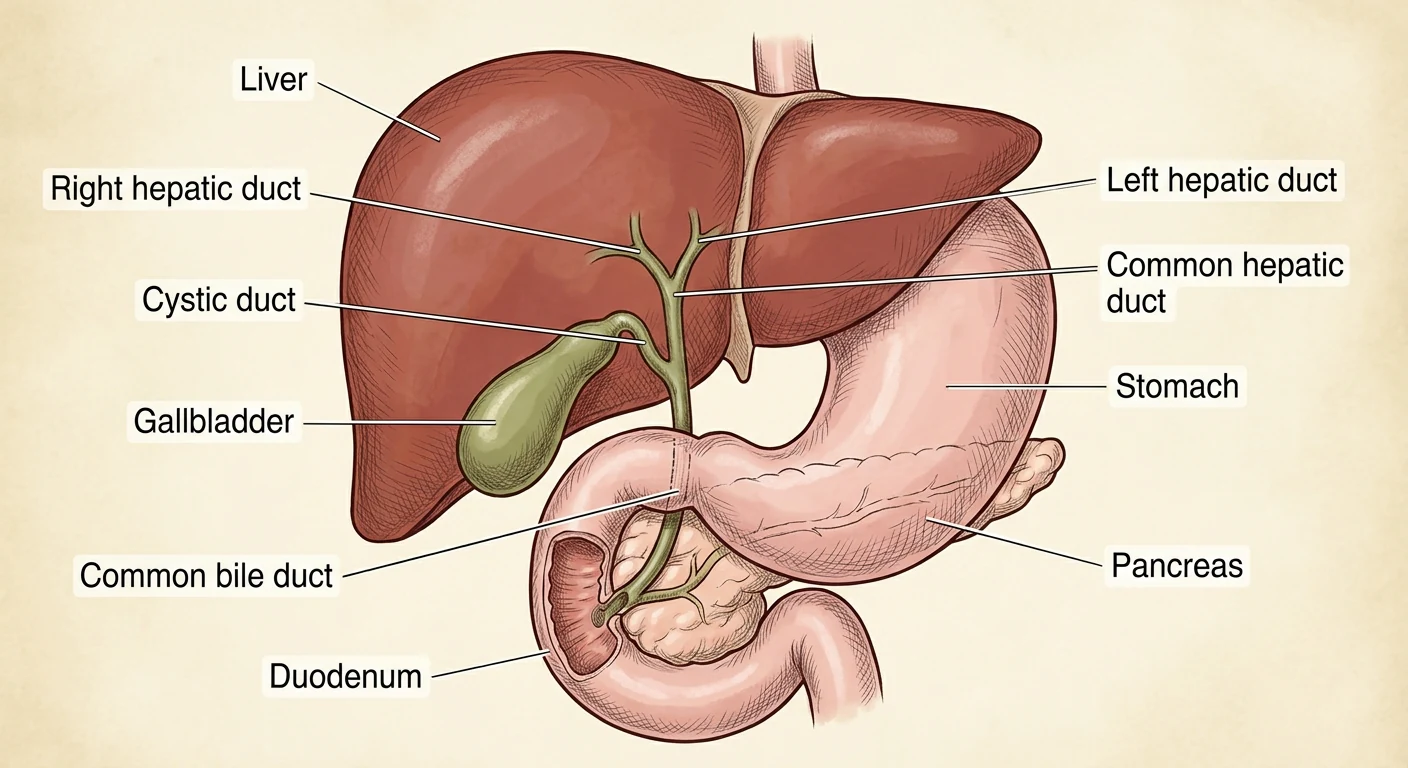
The same rule applies to the arteries (not shown in the figure). The gallbladder has its own artery, the cystic artery. It usually comes off the right hepatic artery, the vessel that feeds the right half of your liver, then runs alongside the cystic duct and spreads out across the surface of the gallbladder. In some people, the cystic artery branches into an anterior and posterior branch (running behind the gallbladder).
Working directly against the gallbladder wall as I clear the space under it of fat and connective tissue keeps the right hepatic artery safe and helps me identify a posterior branch of the cystic artery.
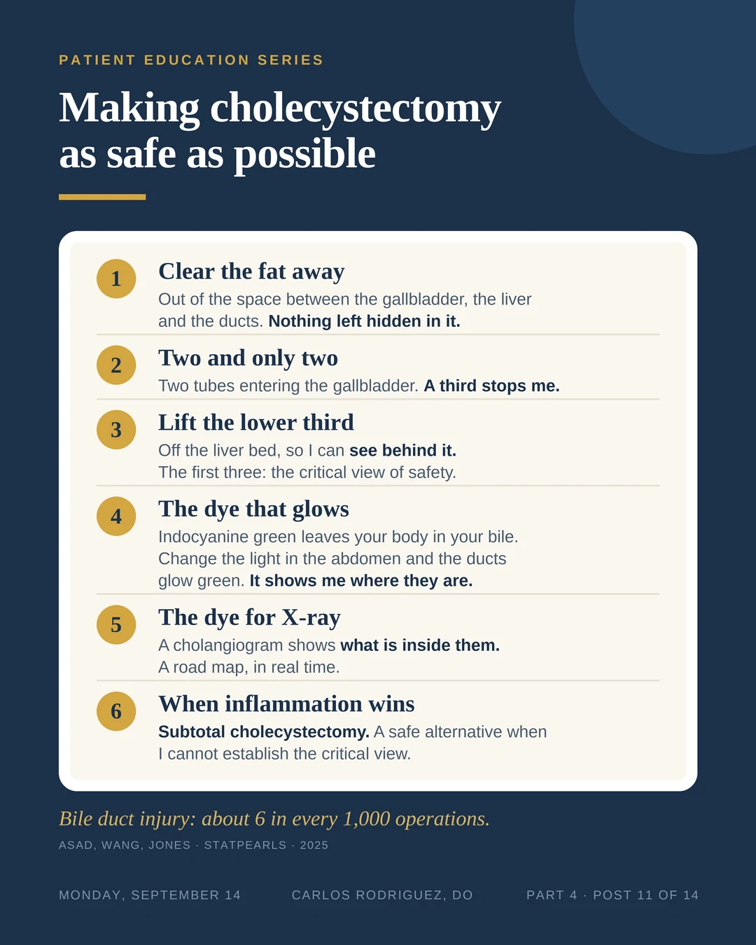
Bile duct injury is the complication every gallbladder surgeon works to prevent. It happens in roughly 6 of every 1,000 gallbladder operations, and about 1 in 1,000 is serious enough to need a reconstructive operation to repair it.¹ Those numbers are small, and they are no comfort to the person it happens to. Everything below is what I do to keep the risk down.
The critical view of safety
Bile ducts almost never get injured through carelessness. They get injured because the anatomy looked like something it was not. Inflammation pulls the structures together and hides them in fat, and the wrong tube can look exactly like the right one.
So surgery uses a rule that does not depend on the tube looking right. It is called the critical view of safety, and it has three parts. All three have to be there before anything is clipped or cut:¹
1. The fat is cleared. All of the fatty tissue comes out of the small space between the gallbladder, the liver and the ducts, until nothing is hidden in it.
2. Two structures, and only two. Exactly two tubes can be seen entering the gallbladder: the cystic duct and the cystic artery. If there is a third thing, the dissection stops until I know what it is.
3. The lower third is off the liver. The bottom of the gallbladder is lifted off its bed on the liver, so I can see behind it.
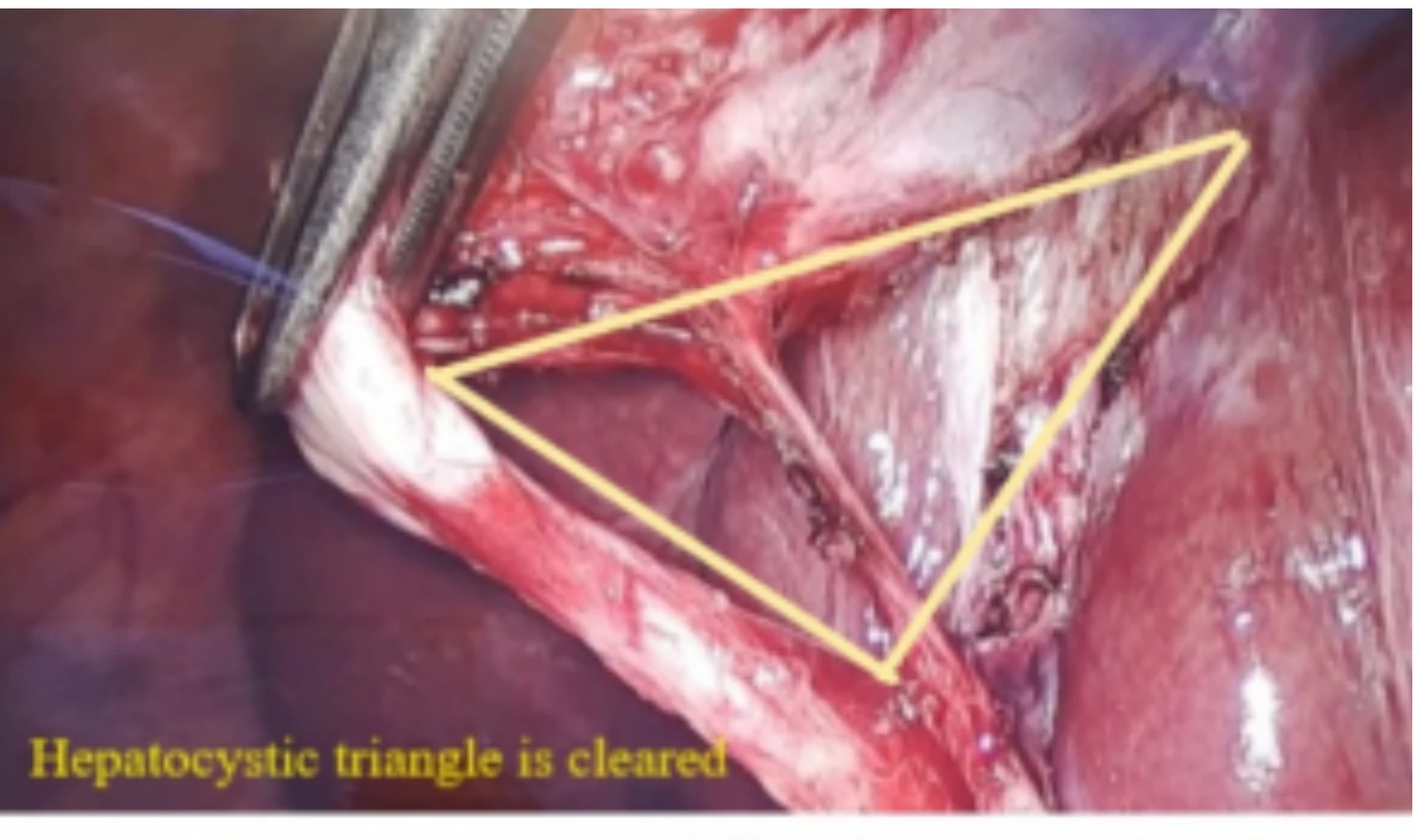
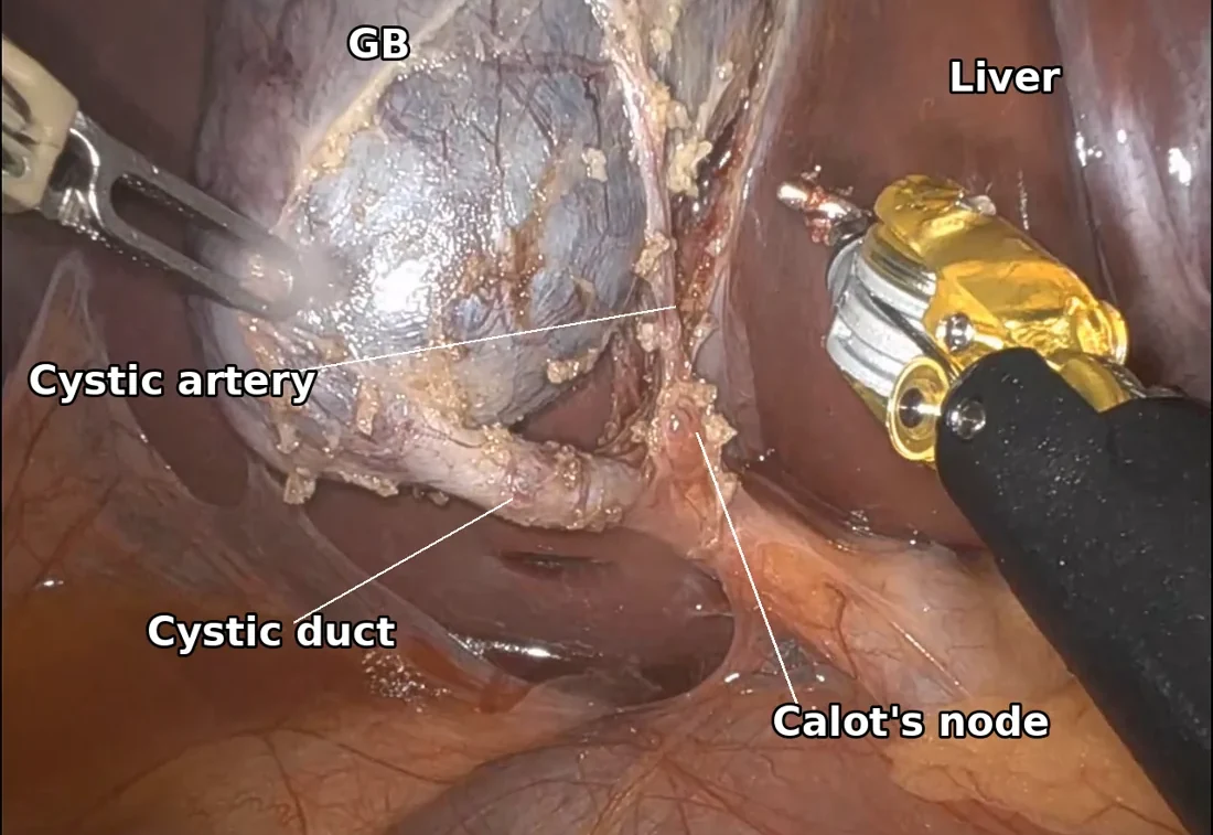
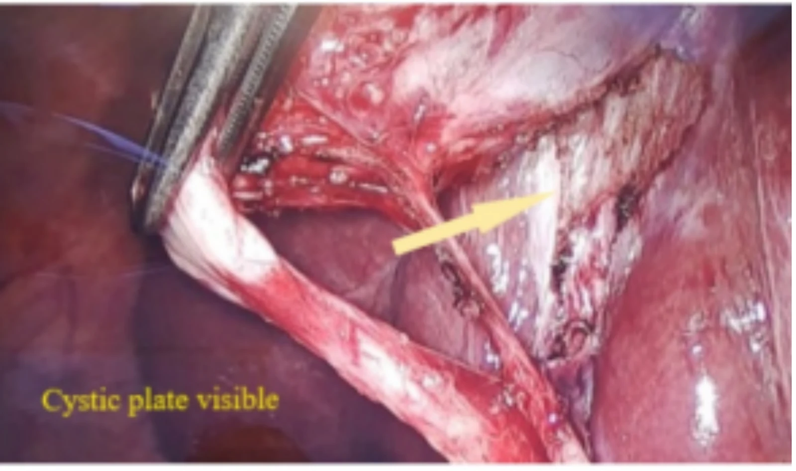
Once those three things are true, the only two structures that can be sitting there are the two that belong to the gallbladder. Nothing else reaches that spot. The rule works by ruling everything else out, rather than by asking me to recognize the right duct on sight.
Nothing gets clipped until I have that view.
The dye that makes the ducts glow
Indocyanine green (ICG) is a dye that has been used in medicine for decades. It goes into a vein before the operation. Your liver pulls it out of your blood and it leaves your body in your bile, which is where I need it. When I switch the camera in your abdomen to a specific setting, your ducts glow green. This is called near-infrared fluorescence cholangiography, and I use it in every case.
I give 5 mg in the pre-op holding area at least 60 minutes before surgery. Other surgeons give different amounts, some a good deal less. I do not think there is a right number.
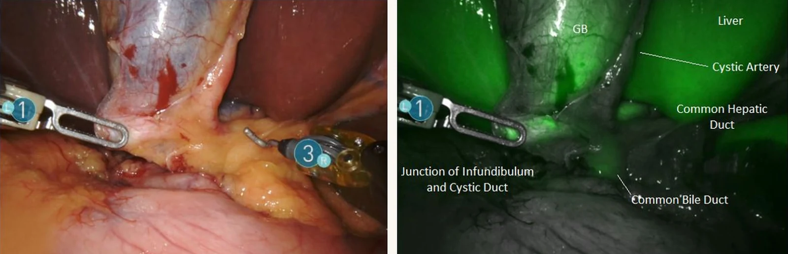
The FALCON trial was an international randomized controlled trial, which means patients were assigned by chance to one approach or the other, so the two groups were comparable. It included 294 patients: 143 had fluorescence imaging and 151 had a standard operation.²
Surgeons using ICG reached the critical view of safety sooner, at an average of 19 minutes 14 seconds against 23 minutes 9 seconds. They found the cystic duct much sooner, 6 minutes 47 seconds against 13 minutes.²
The trial found no difference between the two groups in how long patients stayed in the hospital or in how many had complications after surgery.² One patient developed a rash after the ICG was injected.
FALCON was not built to answer the injury question. Bile duct injury is too rare for 294 patients to show anything. Answering it takes hundreds of thousands of operations, and a study that size came out this year.
Researchers looked at 1,266,024 laparoscopic gallbladder operations done in the United States between 2016 and 2024. About 13 percent used ICG. The two groups were statistically matched on patient and hospital factors, then followed for a year after surgery.³
The patients whose surgeon used ICG had fewer bile duct injuries, 0.25 percent against 0.40 percent. That works out to about 2.5 injuries per 1,000 operations with ICG and about 4 per 1,000 without it. They also needed fewer follow-up procedures on the bile ducts, had fewer operations converted to an open incision, and had fewer complications of any kind in the first 30 days.³
This was not a randomized trial. It looked back at operations that had already happened, which is weaker evidence than FALCON, even with a million more patients in it. The study also excluded robotic operations, which is how I do every one of mine. I expect ICG behaves the same on my platform, but that study did not test it.³
ICG shows me the anatomy earlier, and in the biggest study we have, it came with fewer injured bile ducts. I use it on every case.
Asking for a picture of the ducts
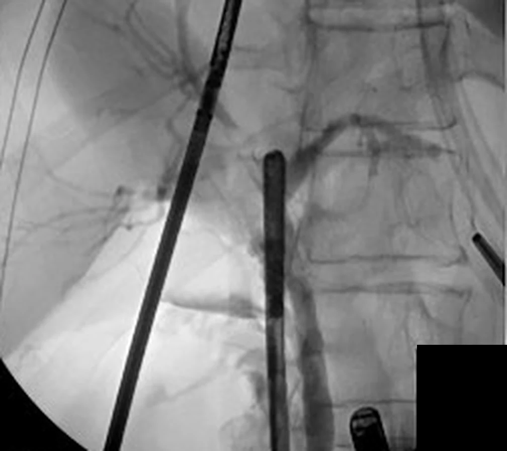
There is a second way to see the bile ducts during surgery, an intraoperative cholangiogram. This one uses a different dye. ICG goes into a vein and glows under a special camera. A cholangiogram uses X-ray contrast, the kind of dye used for a CT scan, and it goes straight into the duct system through a small opening in the cystic duct. An X-ray is then taken on the operating table. It maps the ducts, and it lets me see inside the duct system.
In 2020, five surgical societies published a joint guideline on safe gallbladder surgery. Most of what that document recommends is worded cautiously, because the quality of the underlying evidence is low. It reached agreement on 17 of its 18 questions, and only two recommendations were graded as strong.
One of those two: use imaging of the bile ducts during the operation whenever the anatomy is uncertain or an injury is suspected.⁴
That was 2020. This year SAGES published a guideline on this one question and went further. Its recommendations are conditional, because the certainty of the evidence is still low, but every one of them favors cholangiography: cholangiography over no imaging, cholangiography routinely rather than only in selected cases, and cholangiography over fluorescence with ICG.⁵ A correction published alongside it restates those recommendations in the conclusion, and none of them were reversed. On timing, that guideline was written before the 1.2 million patient study above came out.
The reason for preferring the X-ray contrast over ICG is not that one is better at everything. ICG lights up the outside of the ducts. A cholangiogram fills the ducts with contrast, so it shows what is inside them: a stone sitting in the duct, a branch that ends where it should not, contrast that never reaches the intestine. They answer different questions, so I use both.
The same guideline says the recommendation should be individualized when the blood work is normal, the critical view is definitive, and there is no other reason to take the picture.
Uncertainty was always a reason to shoot a cholangiogram. What changed this year is the case for shooting one when there is no uncertainty.
When inflammation wins
Some gallbladders will not cooperate. The inflammation is dense enough that the cystic duct and artery cannot be safely identified no matter how much time I spend, and the critical view will not be achieved.
One of mine. When the camera went in, there was no gallbladder to see.
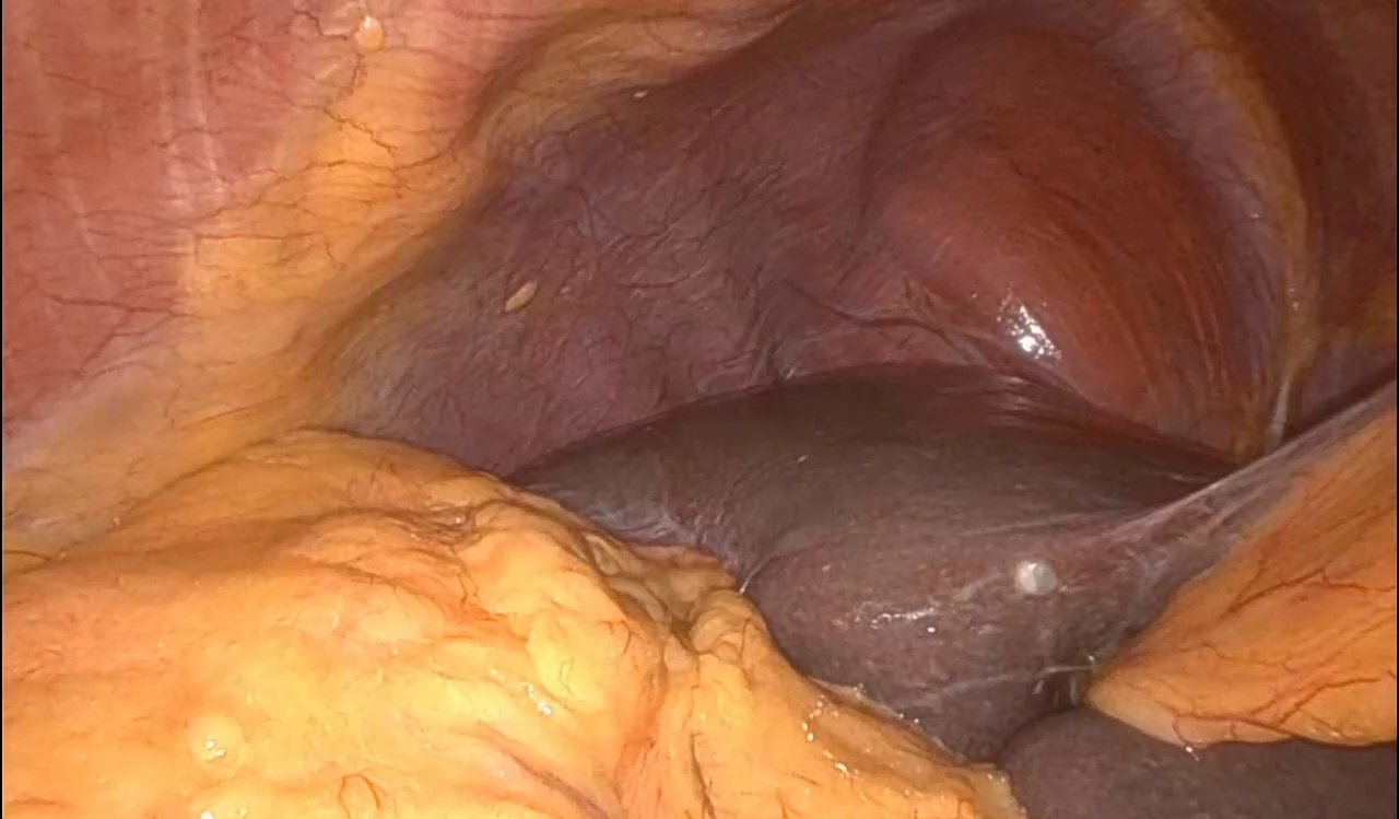
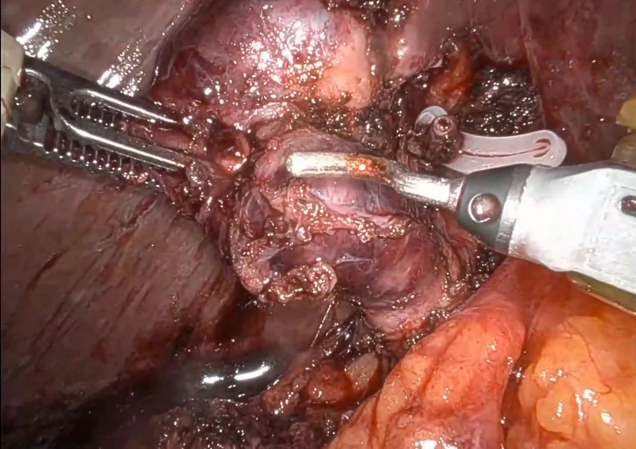
When the triangle is that inflamed or fibrotic, the right answer may be a subtotal cholecystectomy. I take out as much of the gallbladder as I safely can, along with every stone I can find, and leave behind the part that cannot be removed safely.
A review pooling ten studies of difficult gallbladders compared subtotal cholecystectomy with removing the whole organ. Subtotal lowered the risk of common bile duct injury, 0 percent against 1.6 percent.⁶
It raised other risks. Bile leaking afterward, usually from the cystic duct, was about three and a half times more common.⁶
In my opinion, dealing with a cystic duct leak beats dealing with a common bile duct injury.
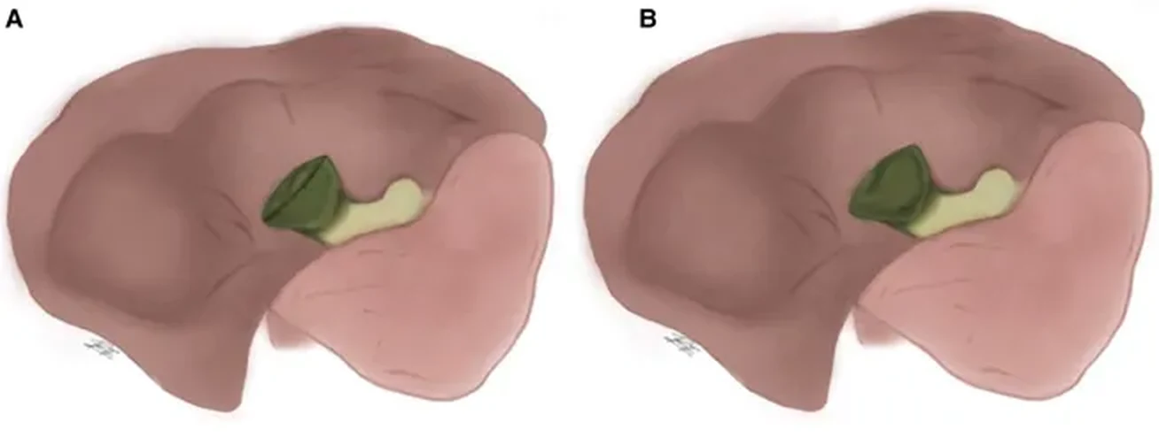
There are two versions of the operation. The reconstituting type closes off the lower end of the gallbladder, which lowers the chance of a leak but leaves a small remnant that can form new stones later. The fenestrating type leaves the gallbladder open, which carries a higher chance of a leak but does not leave a remnant behind to make stones.⁷
What this means for you
If you are having your gallbladder out, feel free to ask:
- Do you use the critical view of safety, and what do you do if you cannot get it?
- Do you use fluorescence imaging, or a cholangiogram, or both?
- How many of these operations do you and this hospital do? In a Swedish registry of 154,934 cholecystectomies, surgeons and hospitals doing fewer of them had more bile duct injuries.⁸
Any surgeon who does this work regularly will have quick, comfortable answers. These are the same three questions I would ask.
If you want to see what surgeons are taught about this, SAGES, the Society of American Gastrointestinal and Endoscopic Surgeons, runs a Safe Cholecystectomy Program built on six steps of its own: get the critical view, expect the anatomy to vary, use imaging of the ducts, pause before cutting anything, stop dissecting when the inflammation is too dense to work through, and call a colleague when the anatomy will not declare itself. The teaching modules and videos are online and open to anyone who creates an account.⁹
This is Part 4 of the gallbladder series. Earlier parts covered the anatomy, how gallstones form, the conditions they cause, the imaging used to find them, and the operation itself.
References
1. Asad U, Wang CF, Jones MW. Laparoscopic Cholecystectomy. In: StatPearls. Treasure Island (FL): StatPearls Publishing; updated July 2, 2025. https://www.ncbi.nlm.nih.gov/books/NBK448145/
2. van den Bos J, Schols RM, Boni L, et al. Near-infrared fluorescence cholangiography assisted laparoscopic cholecystectomy (FALCON): an international multicentre randomized controlled trial. Surg Endosc. 2023;37(6):4574-4584. https://pubmed.ncbi.nlm.nih.gov/36849564/
3. Neel NC, Van Doosselaere L, Thareja NS, et al. Indocyanine Green Fluorescent Cholangiography During Laparoscopic Cholecystectomy and Bile Duct Injury. JAMA Surg. 2026;161(9):927-934. https://pubmed.ncbi.nlm.nih.gov/42485022/
4. Brunt LM, Deziel DJ, Telem DA, et al. Safe Cholecystectomy Multi-society Practice Guideline and State of the Art Consensus Conference on Prevention of Bile Duct Injury During Cholecystectomy. Ann Surg. 2020;272(1):3-23. https://pubmed.ncbi.nlm.nih.gov/32404658/
5. Kumar SK, Shehata DG, Cetrulo LN, et al. SAGES guidelines for the use of intraoperative imaging of the common bile duct. Surg Endosc. 2025;39(11):7091-7102. https://pubmed.ncbi.nlm.nih.gov/40935939/ (Correction: Surg Endosc. 2025;39(11):7854-7855. https://pubmed.ncbi.nlm.nih.gov/41044285/)
6. Koo SS, Krishnan RJ, Ishikawa K, et al. Subtotal vs total cholecystectomy for difficult gallbladders: A systematic review and meta-analysis. Am J Surg. 2023;229:145-150. https://pubmed.ncbi.nlm.nih.gov/38168604/
7. Strasberg SM, Pucci MJ, Brunt LM, Deziel DJ. Subtotal Cholecystectomy, "Fenestrating" vs "Reconstituting" Subtypes and the Prevention of Bile Duct Injury. J Am Coll Surg. 2016;222(1):89-96. https://pubmed.ncbi.nlm.nih.gov/26521077/
8. Blohm M, Sandblom G, Enochsson L, et al. Relationship between surgical volume and outcomes in elective and acute cholecystectomy: nationwide, observational study. Br J Surg. 2023;110(3):353-361. https://pubmed.ncbi.nlm.nih.gov/36422988/
9. Society of American Gastrointestinal and Endoscopic Surgeons. The SAGES Safe Cholecystectomy Program: Strategies for Minimizing Bile Duct Injuries. https://www.sages.org/safe-cholecystectomy-program/