Thank you for following all 14 posts in the gallbladder series. Enough about stones. Let's talk about hernias.
For the next 14 weeks, this series covers inguinal hernias, the hernias that form in the inguinal region. Hernia repair in the inguinal region is one of the most common operations in the world, with more than 20 million done each year.¹ I will follow the same order I used for the gallbladder: anatomy first, then what goes wrong, how I repair it, and what recovery looks like.
This first post is a little longer than usual. The inguinal region forms differently in men and women, so I cover both. Part 1 is for patients. Part 2 is for medical students and residents.
Part 1: How the inguinal region forms
The anatomy of the inguinal region is complex. It makes the most sense if you start before birth and slowly build the anatomy one layer at a time. The video below shows how the inguinal region is formed.
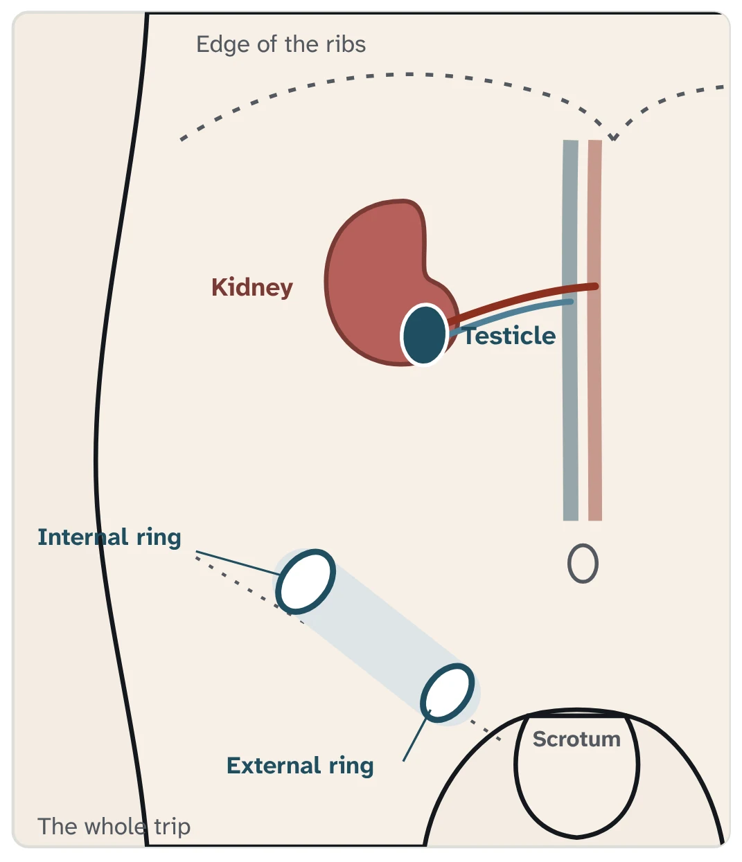
The testicle starts high in the abdomen
A baby boy's testicles do not start in the scrotum. Each one forms inside the abdomen, high in the back, beside the kidney.² The artery that feeds it branches off the aorta, the body's main artery, near the kidneys. When the testicle moves down, its blood vessels come with it.²
It moves down in two stages
Between about 8 and 15 weeks of pregnancy, a strand of tissue called the gubernaculum holds the testicle near the inguinal region while the baby grows. Between about 25 and 35 weeks, the testicle follows the gubernaculum through the wall of the abdomen and into the scrotum.³
The tunnel: the inguinal canal
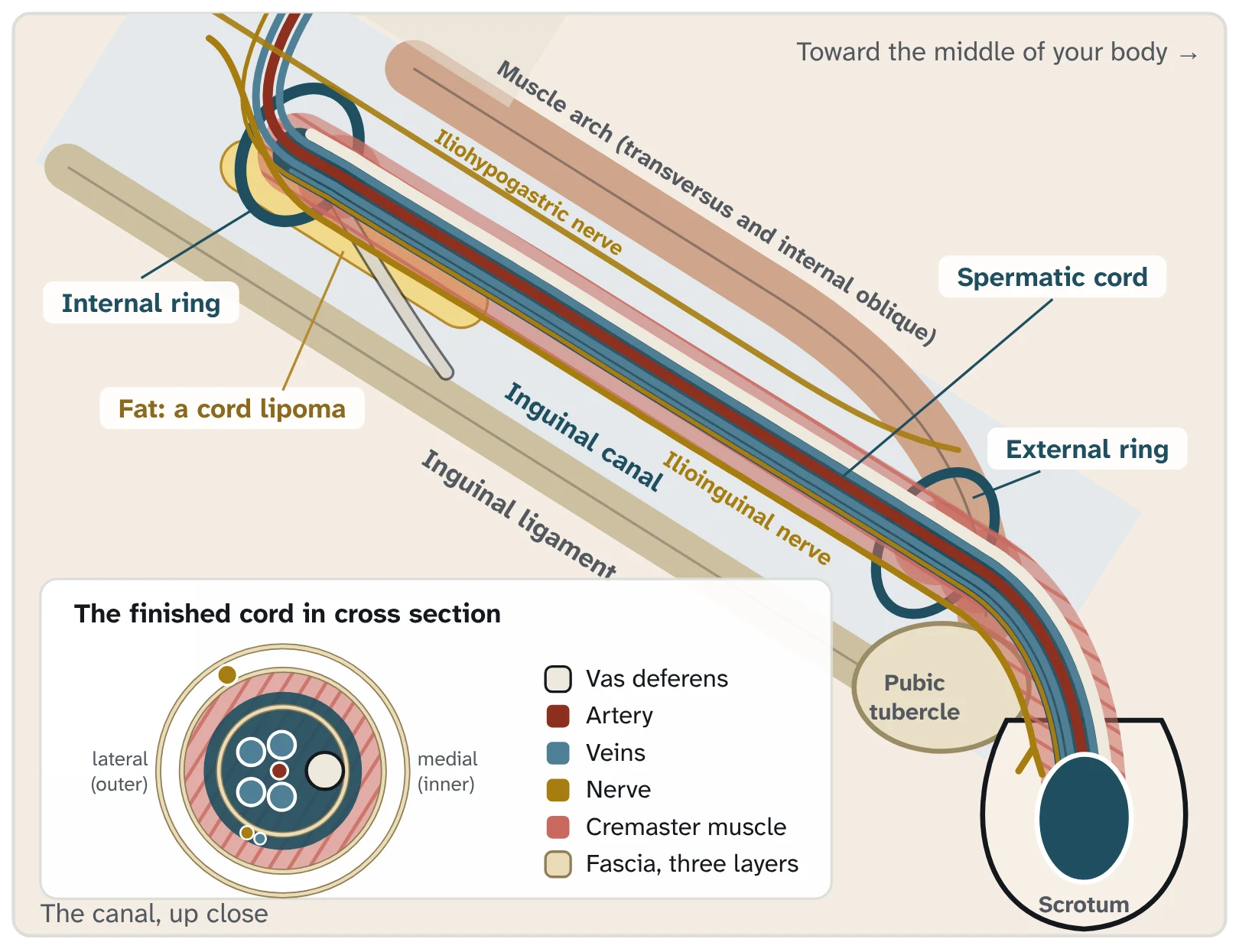
The testicle leaves the abdomen through a short tunnel in the lower abdominal wall called the inguinal canal. It is about 4 to 6 centimeters long, roughly 1.5 to 2.5 inches. It has an opening at each end: the internal ring on the inside and the external ring just above the pubic bone.⁴
Everything the testicle needs passes through the tunnel in one bundle, the spermatic cord. The cord holds the tube that carries sperm (the vas deferens), the blood vessels of the testicle and small nerves. A thin muscle called the cremaster and three thin layers of tissue wrap around them.⁴,⁵
Women have the same tunnel
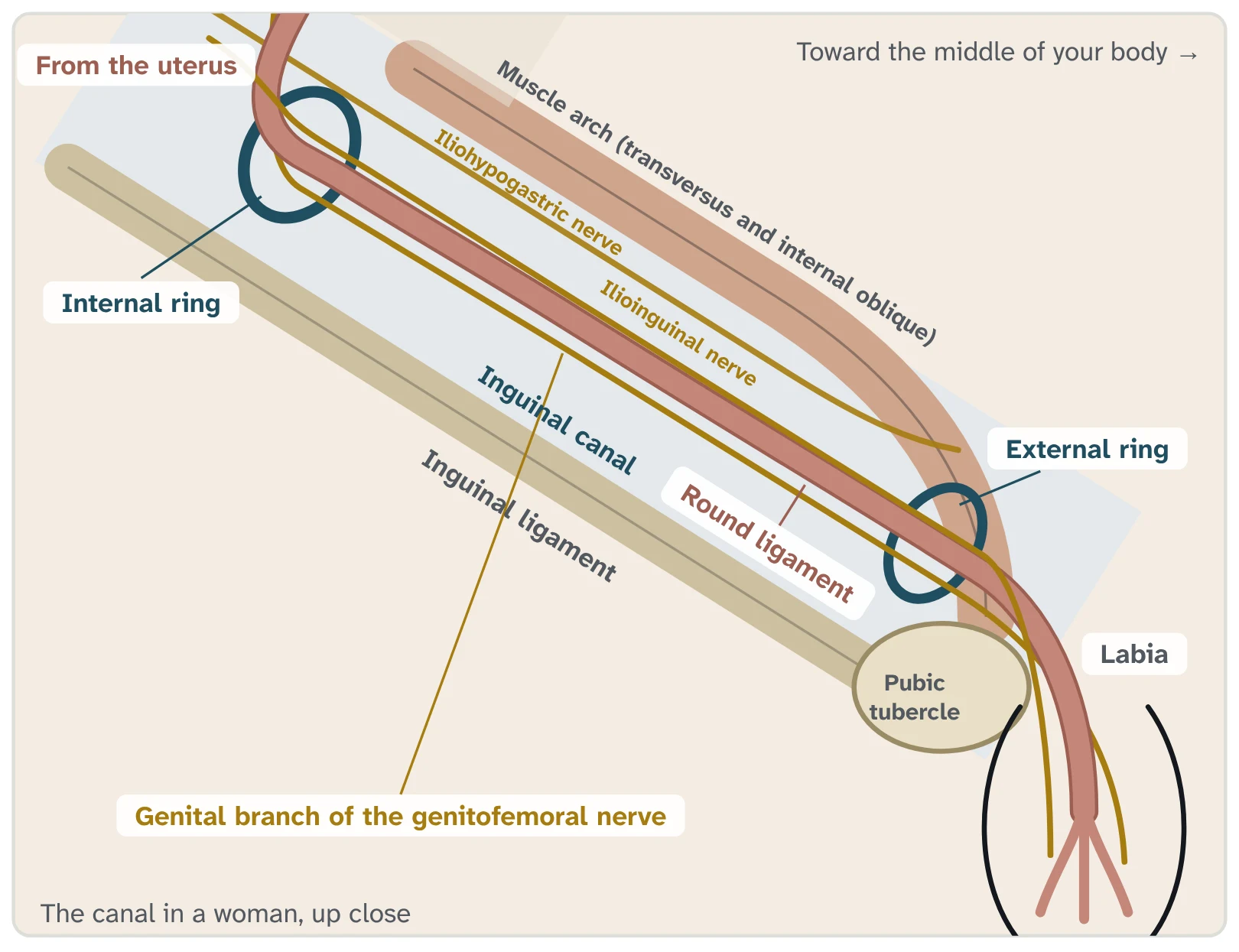
In a woman, the inguinal canal carries the round ligament, a band of tissue that runs from the uterus, through the canal, to the labia. It forms from the same strand of tissue that guides the testicle down in a boy.⁶ Before birth, this strand grows from the inguinal region toward the ovary. On its way, it crosses the tube that will become the uterus and attaches to it.⁶,⁷ That is why the round ligament starts at the top corner of the uterus.
The round ligament helps keep the uterus tipped forward during pregnancy. Other, stronger ligaments do most of the work of holding the uterus in place.⁶ Later in this series, I will explain why I usually divide the round ligament during a robotic repair.
Inguinal hernias happen in women too. A later post covers why they are often missed.
Why this matters for hernias
Before birth, a thin pouch of the lining of the abdomen goes down the tunnel with the testicle. It should close. In about 1 adult in 8, it stays open, usually with no symptoms.⁸ People with an open pouch are about four times more likely to need a hernia repair later.⁹
The tunnel itself never closes, because the cord or the round ligament passes through it for life. That leaves a natural weak spot in the wall of the abdomen. Inguinal hernias form in and around this tunnel.
Next week, I will show you the same area from the inside, the way I see it during robotic surgery. The full animation, including the two types of inguinal hernia and how I repair them through an open incision, is on my inguinal hernia page.
Part 2: Resident Pearls
This part is longer than usual. It covers the anatomy you will encounter during an open repair.
Resident Pearls
1. Boundaries. The anterior wall is the external oblique aponeurosis, with internal oblique contributing laterally. The posterior wall is transversalis fascia, with the conjoint tendon medially. Surgeons call this posterior wall the floor of the canal. The cephalad border is the arching fibers of internal oblique and transversus abdominis. The caudal border is the inguinal ligament, with the lacunar ligament medially.¹⁰,¹¹
2. The rings, and the line between direct and indirect. The deep ring is a defect in transversalis fascia about 1 cm above the inguinal ligament and lateral to the inferior epigastric vessels. The superficial ring is a V-shaped opening in the external oblique aponeurosis, above and lateral to the pubic tubercle.¹⁰ An indirect hernia leaves lateral to the epigastric vessels, through the deep ring and inside the cord. A direct hernia comes through Hesselbach's triangle, medial to them.⁴,¹¹
3. Each covering maps to a wall layer. External spermatic fascia comes from the external oblique aponeurosis, the cremaster and cremasteric fascia from the internal oblique, and the internal spermatic fascia from transversalis fascia.⁵ The peritoneum that descends with the testis becomes the tunica vaginalis.¹² Lie and Hutson argue the cremaster differentiates from the gubernacular tip and is not a passive extension of the internal oblique. Most of that evidence is from rodents, so the classic description still holds at operation.¹³
4. What sits where inside the cord. Konschake and colleagues confirmed each nerve with immunohistochemistry.¹⁴ The ilioinguinal nerve lies between the external spermatic and cremasteric fascia. It travels with the cord but is not a true cord content. The genital branch of the genitofemoral nerve runs in the cremasteric layer with the cremasteric vein, the "blue line." Opening the internal spermatic fascia exposes the vas, the testicular artery and the pampiniform plexus.¹⁴ A cord lipoma arises from preperitoneal fat in this same compartment and is classified as a lateral hernia.⁵
5. Iliohypogastric nerve. It runs between the internal and external oblique, above the cord, and pierces the external oblique aponeurosis a mean of 3.8 cm above the superficial ring, with a range of 2.5 to 5.5 cm.¹⁵
6. Ilioinguinal nerve. It does not enter through the deep ring. It pierces the internal oblique lateral to the ring, enters the canal from the side and lies under the external oblique aponeurosis on the front of the cord.¹⁴,¹⁵ In 100 cadaver dissections it lay in front of the cord in 79% and was absent in 7%.¹⁶ At the superficial ring it exits lateral and superficial to the cord.¹⁴
7. Genital branch of the genitofemoral nerve. It enters through the deep ring and stays inside the cord. In 116 cadaver sides, it ran between the cremaster fibers and the internal spermatic fascia, most often along the inferior fibers (59%), and only 3% ran outside the cord.¹⁷ In 244 Lichtenstein repairs, surgeons identified the iliohypogastric nerve in 95%, the ilioinguinal in 98% and the genital branch in only 21%.¹⁸ I open the cremaster as part of my dissection, and I don't think I have ever seen this nerve.
8. Expect variation. In 110 open repairs, the ilioinguinal and iliohypogastric nerves both followed the textbook course in only 42%. Variants included a single shared trunk, an absent nerve and an ilioinguinal nerve running outside the aponeurosis.¹⁹ The international guidelines recommend that open surgery focus on recognizing the nerves.¹
9. The female canal. The round ligament is about 10 to 12 cm long, runs from the uterine cornu through the canal to the labium majus and mons pubis, and carries the Sampson artery.⁶ The genital branch travels with it.¹⁴ The female counterpart of the processus vaginalis is the canal of Nuck. If it stays open, it can present as a cyst or a hernia, and a hernia coexists in up to 40% of cysts.²⁰
10. Embryology of descent. The gonad forms on the urogenital ridge, which is why the testicular artery arises from the aorta just below the renal arteries.²,¹² Descent is biphasic. In the transabdominal phase (about 8 to 15 weeks), INSL3 from Leydig cells swells the gubernaculum and anchors the testis near the future canal, while testosterone causes the cranial suspensory ligament to regress. The inguinoscrotal phase (about 25 to 35 weeks) is androgen dependent. The genitofemoral nerve and CGRP model of that phase comes from animal work, with little direct human evidence.³
11. The patent processus vaginalis. In 599 adults having laparoscopy for other reasons, about 1 in 8 had an asymptomatic patent processus: right-sided in 59%, left in 29% and bilateral in 12%. Its prevalence did not rise with age, and the authors concluded that adult indirect hernia is congenital.⁸ On follow-up, a patent processus carried about four times the risk of a later hernia repair.⁹
References
1. HerniaSurge Group. International guidelines for groin hernia management. Hernia. 2018;22(1):1-165. https://pubmed.ncbi.nlm.nih.gov/29330835/
2. Tiwana MS, Leslie SW. Anatomy, Abdomen and Pelvis: Testes. StatPearls [Internet]. Treasure Island (FL): StatPearls Publishing. https://www.ncbi.nlm.nih.gov/books/NBK470201/
3. Hutson JM, Li R, Southwell BR, Newgreen D, Cousinery M. Regulation of testicular descent. Pediatr Surg Int. 2015;31(4):317-325. https://pubmed.ncbi.nlm.nih.gov/25690562/
4. Tuma F, Lopez RA, Varacallo MA. Anatomy, Abdomen and Pelvis: Inguinal Region (Inguinal Canal). StatPearls [Internet]. Treasure Island (FL): StatPearls Publishing; updated July 24, 2023. https://www.ncbi.nlm.nih.gov/books/NBK470204/
5. Köckerling F, Schug-Pass C. Spermatic cord lipoma: a review of the literature. Front Surg. 2020;7:39. https://pubmed.ncbi.nlm.nih.gov/32793626/
6. Chaudhry SR, Chaudhry K. Anatomy, Abdomen and Pelvis: Uterus Round Ligament. StatPearls [Internet]. Treasure Island (FL): StatPearls Publishing; updated July 24, 2023. https://www.ncbi.nlm.nih.gov/books/NBK499970/
7. Acién P, Sánchez del Campo F, Mayol MJ, Acién M. The female gubernaculum: role in the embryology and development of the genital tract and in the possible genesis of malformations. Eur J Obstet Gynecol Reprod Biol. 2011;159(2):426-432. https://pubmed.ncbi.nlm.nih.gov/21831509/
8. van Wessem KJP, Simons MP, Plaisier PW, Lange JF. The etiology of indirect inguinal hernias: congenital and/or acquired? Hernia. 2003;7(2):76-79. https://pubmed.ncbi.nlm.nih.gov/12820028/
9. van Veen RN, van Wessem KJP, Halm JA, et al. Patent processus vaginalis in the adult as a risk factor for the occurrence of indirect inguinal hernia. Surg Endosc. 2007;21(2):202-205. https://pubmed.ncbi.nlm.nih.gov/17122977/
10. Inguinal canal. Radiopaedia.org. https://radiopaedia.org/articles/inguinal-canal
11. Sugumar K, Gupta M. Anatomy, Abdomen and Pelvis: Inguinal Ligament (Crural Ligament, Poupart Ligament). StatPearls [Internet]. Treasure Island (FL): StatPearls Publishing; updated January 30, 2024. https://www.ncbi.nlm.nih.gov/books/NBK542321/
12. Titi-Lartey OA, Khan YS. Embryology, Testicle. StatPearls [Internet]. Treasure Island (FL): StatPearls Publishing; updated April 24, 2023. https://www.ncbi.nlm.nih.gov/books/NBK557763/
13. Lie G, Hutson JM. The role of cremaster muscle in testicular descent in humans and animal models. Pediatr Surg Int. 2011;27(12):1255-1265. https://pubmed.ncbi.nlm.nih.gov/22038274/
14. Konschake M, Zwierzina M, Moriggl B, et al. The inguinal region revisited: the surgical point of view. Hernia. 2020;24(4):883-894. https://pubmed.ncbi.nlm.nih.gov/31776877/
15. Wijsmuller AR, Lange JFM, Kleinrensink GJ, et al. Nerve-identifying inguinal hernia repair: a surgical anatomical study. World J Surg. 2007;31(2):414-420. https://pubmed.ncbi.nlm.nih.gov/17180560/
16. Ndiaye A, Diop M, Ndoye JM, et al. Anatomical basis of neuropathies and damage to the ilioinguinal nerve during repairs of groin hernias (about 100 dissections). Surg Radiol Anat. 2007;29(8):675-681. https://pubmed.ncbi.nlm.nih.gov/17985072/
17. Liu WC, Chen TH, Shyu JF, et al. Applied anatomy of the genital branch of the genitofemoral nerve in open inguinal herniorrhaphy. Eur J Surg. 2002;168(3):145-149. https://pubmed.ncbi.nlm.nih.gov/12182238/
18. Bischoff JM, Aasvang EK, Kehlet H, Werner MU. Does nerve identification during open inguinal herniorrhaphy reduce the risk of nerve damage and persistent pain? Hernia. 2012;16(5):573-577. https://pubmed.ncbi.nlm.nih.gov/22782363/
19. al-Dabbagh AKR. Anatomical variations of the inguinal nerves and risks of injury in 110 hernia repairs. Surg Radiol Anat. 2002;24(2):102-107. https://pubmed.ncbi.nlm.nih.gov/12197017/
20. Kohlhauser M, Pirsch JV, Maier T, Viertler C, Fegerl R. The cyst of the canal of Nuck: anatomy, diagnostic and treatment of a very rare diagnosis. A case report of an adult woman and narrative review of the literature. Medicina (Kaunas). 2022;58(10):1353. https://pubmed.ncbi.nlm.nih.gov/36295514/
Dr. Rodriguez served as a speaker and educator for Intuitive Surgical through June 2025. He has no current financial relationship with the company.