Last week, I showed how the inguinal region forms before birth. This week, we look at the same area the way I see it during robotic surgery.
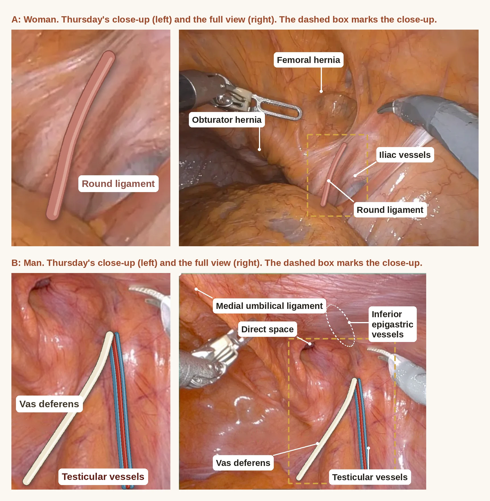
On Thursday, I posted two close-up photos on LinkedIn and asked which one was a man and which one was a woman. Photo A is a woman. Photo B is a man (Figure 1). This post explains how to tell them apart.
Part 1 is for patients. Part 2 is for medical students and residents.
Part 1: The view from inside
What the camera sees
During a robotic hernia repair, the camera goes in through a small incision higher on the abdomen and looks down toward the inguinal region. Figures 1A and 1B are good examples of my first view. A thin, shiny inner lining, the peritoneum, covers the inside of the abdominal wall.
Through that lining, you can make out the vessels and cords that run toward the internal ring. The internal ring is the inside opening of the inguinal canal, the tunnel from last week's post.
The landmark I find first
The first landmarks I see are the inferior epigastric vessels (Figure 1B). They are an artery and a vein that run up the inside of the abdominal wall. The internal ring sits just to the outer side of them.¹
These vessels mark the line between the two common kinds of inguinal hernia. A hernia that comes out through the internal ring is called an indirect hernia.¹ A hernia that pushes through the back wall of the canal, on the inner side of the vessels, is called a direct hernia.¹ I will show both kinds in a later post.
In a man, you can still see the path the testicle took
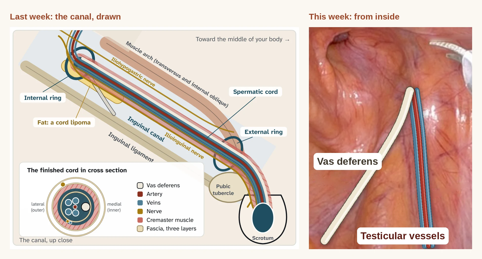
Last week's video showed the testicle starting high in the abdomen, beside the kidney, then moving down and out through the inguinal canal. It brought its blood supply and the vas deferens along with it. From inside, you can still see that path (Figure 2).
The testicular vessels. These are the artery and veins that supply the testicle. They start high in the abdomen, near the kidney, where the testicle started. They come down to the ring on the outer side.
The vas deferens. This is the tube that carries sperm. It comes up out of the pelvis to the ring on the inner side.
The two meet at the internal ring and leave the abdomen together as the spermatic cord, the same cord you saw running through the canal in last week's video. From inside, they form a narrow V with its point at the ring. Studies on cadavers measured that angle at about 40 to 50 degrees.²,³
The space inside that V matters during surgery. Surgeons call it the triangle of doom. The large blood vessels to the leg run underneath it.² Surgeons keep stitches and tacks out of that space.
In a woman, the ovary stayed in the pelvis
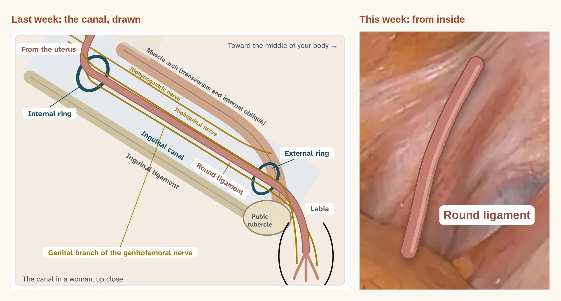
The women's video showed the ovary moving down too, then stopping in the pelvis. Its blood vessels stay in the pelvis with it. The only structure that reaches the internal ring is the round ligament (Figure 3), a cord of tissue about 10 to 12 cm long. It starts at the top corner of the uterus, crosses the pelvis, passes through the internal ring and runs down the canal.⁴ It grows from the same tissue that guides the testicle down in a boy.⁴ Since a woman has no spermatic cord, the triangle of doom has only an inner edge: the round ligament.
That is the difference you were asked to spot on Thursday. In a man, two structures meet at the ring. In a woman, there is one.
The pouch, from inside
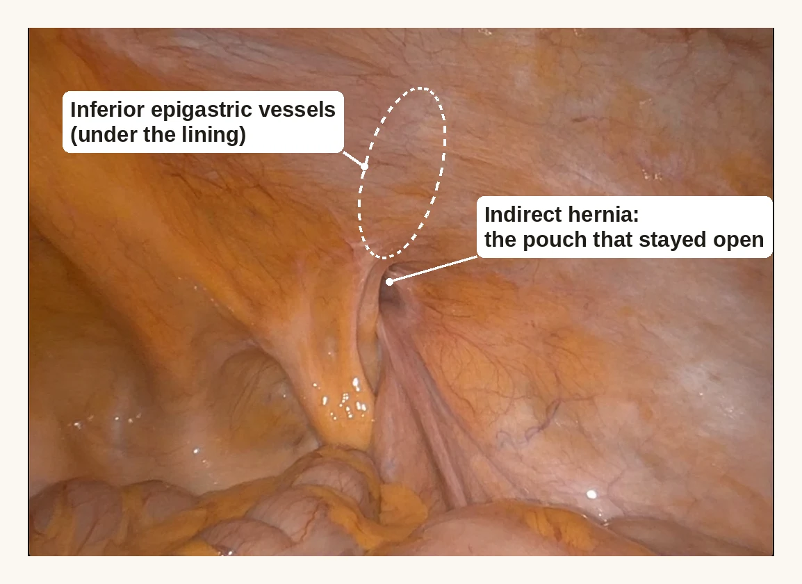
Last week's video also showed a thin pouch of lining that follows the testicle down. It normally closes after birth. From inside, a closed pouch leaves a smooth surface over the ring. A pouch that stays open shows up as an opening at the ring.
Figure 4 shows what that looks like in a man. The pouch never closed, and it became an indirect inguinal hernia. Below the opening, the vas deferens and the testicular vessels form the V of the triangle of doom.
Two more openings nearby
Two more openings sit close to the internal ring. The femoral opening sits just below the inguinal region. The obturator opening sits deeper in the pelvis. When they are normal, the shiny inner layer of the abdomen hides these openings. When a hernia forms in one of them, you can see it from inside. The woman in Figure 1A had both a femoral hernia and an obturator hernia. I will cover both in a later post.
This matters most for women. Women have far fewer inguinal hernias than men, but they get femoral hernias more often. In the Swedish Hernia Register, only 8% of hernia repairs in this region were done in women.⁵ Yet women had 2,490 of the 3,980 femoral hernia repairs, nearly two out of three.⁶ Femoral hernias in women are also missed. When women needed a second operation after an inguinal hernia repair, surgeons found a femoral hernia 41.6% of the time, compared with 4.6% in men.⁵ Femoral hernias are also more likely to become emergencies. In the same register, 40.6% of women with a femoral hernia needed emergency surgery, compared with 28.1% of men.⁶
For these reasons, international guidelines suggest that women have their repair done from the inside when the surgeon has that expertise, partly so a femoral hernia is not missed.⁷
Why this matters for hernias
Figure 4 shows a pouch that never closed and became a hernia. An open pouch is common. Surgeons in the Netherlands looked at the internal ring on both sides in 599 adults who were having laparoscopic surgery for other reasons. About 1 in 8 (12%) had an open pouch. Over the next five years, those patients were four times more likely to need a hernia repair than patients whose pouch had closed.⁸
Inguinal hernias are far more common in men. A study from Oxford, England estimated that 27% of men will have an inguinal hernia repair at some point in life.⁹ That is more than 1 in 4 men. For women, the estimate was 3%.⁹
Because the camera can see both sides, international guidelines recommend checking the other side during this type of repair, with the patient's consent.⁷
Next week, I will explain how an inguinal hernia forms, and why one heavy lift is rarely the cause.
Part 2: Resident Pearls
Resident Pearls
1. The same line, seen from behind. Last week's Pearl 2 drew the line between direct and indirect hernias from the anterior view. An indirect sac enters the canal through the deep ring, and a direct sac comes through the posterior wall.¹ The defect is named by where it sits relative to the epigastric vessels.
2. Three folds orient you. From the midline out: the median umbilical ligament (urachus), the medial umbilical ligament (obliterated umbilical artery) and the lateral umbilical fold, which carries the inferior epigastric vessels. They divide the lower anterior wall into the supravesical, medial inguinal and lateral inguinal fossae. Direct hernias come through the medial inguinal fossa. Indirect hernias come through the lateral inguinal fossa at the deep ring.
3. In a man, the vas and the testicular vessels make a V. The vas runs medially from the pelvis and the testicular vessels come down laterally. In cadavers, O'Malley measured the angle at 38.6 degrees on the right and 48.6 degrees on the left, with each side taken from different cadavers.² Gupta measured 43.5 degrees.³
4. The tissue over the vas is thin. O'Malley measured the tissue around the ring at about 0.2 mm over the vas and about 2.2 mm lateral to the testicular vessels.² Expect that when you dissect the peritoneum off the vas.
5. The apex of the V is the apex of the triangle of doom. The external iliac vessels lie beneath it, which is why fixation stays out of this space.² I cover the triangle of doom and the triangle of pain in IH 10, when we raise the flap.
6. The female ring has no V. The round ligament is the only structure entering the deep ring.⁴ Without a vas and testicular vessels to mark the apex, use the inferior epigastric vessels and the round ligament together to find the ring.
7. An open processus is a diagnosis made from this view. Van Veen studied 599 consecutive adults (189 men) having transperitoneal laparoscopy for other reasons, with the deep ring inspected on both sides.⁸ At a mean follow up of 5.5 years, 337 remained in the study. Twelve percent had a patent processus vaginalis. Hernia repair occurred in 12% of the patent group against 3% of the obliterated group, about four times the risk, and prevalence did not change with age.⁸ HerniaSurge recommends inspecting the contralateral side during TAPP after consent, and does not suggest it during unilateral TEP.⁷
8. The intact view does not show the whole myopectineal orifice. Fruchaud's myopectineal orifice includes the indirect, direct and femoral spaces. Stoppa's giant preperitoneal mesh repair was built on covering the whole orifice with wide overlap.¹⁰ Daes and Felix described a critical view of the myopectineal orifice for laparoscopic repair.¹¹ Raise the flap far enough to see all of it before mesh goes in.
9. Learn the critical view from vetted video. Huynh reviewed the 50 most viewed YouTube videos of laparoscopic inguinal hernia repair against the Daes and Felix criteria. Only 16% met all nine, and 46% showed unsafe technique.¹² Use society endorsed video. One I recommend: Dr. David Lourié, Robotic Inguinal Hernia Repair (YouTube, 2020), https://youtu.be/AdlNAr-9JCc
10. A normal ring does not rule out a lipoma of the cord or round ligament. Lilly and Arregui found cord lipomas in 22.5% of 280 repairs, including nine in women.¹³ These lipomas come from retroperitoneal fat outside the internal spermatic fascia, protrude through the ring lateral to the cord and are generally not visible from inside until reduced.¹³ Some cause hernia symptoms with no peritoneal defect.¹³ If the patient had a bulge and the ring looks normal, reduce the preperitoneal fat and look again.
11. In women, think femoral. HerniaSurge suggests laparoendoscopic repair in women when expertise is available, to lower the risk of chronic pain and avoid missing a femoral hernia.⁷ The same guidelines list female sex and femoral hernia as risk factors for incarceration and strangulation.⁷
References
1. Tuma F, Lopez RA, Varacallo MA. Anatomy, Abdomen and Pelvis: Inguinal Region (Inguinal Canal). In: StatPearls. Treasure Island (FL): StatPearls Publishing; updated July 24, 2023. https://www.ncbi.nlm.nih.gov/books/NBK470204/
2. O'Malley KJ, Monkhouse WS, Qureshi MA, Bouchier-Hayes DJ. Anatomy of the peritoneal aspect of the deep inguinal ring: implications for laparoscopic inguinal herniorrhaphy. Clin Anat. 1997;10(5):313-7. https://pubmed.ncbi.nlm.nih.gov/9283728/
3. Gupta R, Gupta T, Kumari C, Aggarwal A, Sahni D. Revisiting the surgical anatomy of the triangle of doom and the triangle of pain. Clin Anat. 2022;35(7):899-905. https://pubmed.ncbi.nlm.nih.gov/35426468/
4. Chaudhry SR, Chaudhry K. Anatomy, Abdomen and Pelvis: Uterus Round Ligament. In: StatPearls. Treasure Island (FL): StatPearls Publishing; updated July 24, 2023. https://www.ncbi.nlm.nih.gov/books/NBK499970/
5. Koch A, Edwards A, Haapaniemi S, Nordin P, Kald A. Prospective evaluation of 6895 groin hernia repairs in women. Br J Surg. 2005;92(12):1553-8. https://pubmed.ncbi.nlm.nih.gov/16187268/
6. Dahlstrand U, Wollert S, Nordin P, Sandblom G, Gunnarsson U. Emergency femoral hernia repair: a study based on a national register. Ann Surg. 2009;249(4):672-6. https://pubmed.ncbi.nlm.nih.gov/19300219/
7. HerniaSurge Group. International guidelines for groin hernia management. Hernia. 2018;22(1):1-165. https://pubmed.ncbi.nlm.nih.gov/29330835/
8. van Veen RN, van Wessem KJP, Halm JA, Simons MP, Plaisier PW, Jeekel J, Lange JF. Patent processus vaginalis in the adult as a risk factor for the occurrence of indirect inguinal hernia. Surg Endosc. 2007;21(2):202-5. https://pubmed.ncbi.nlm.nih.gov/17122977/
9. Primatesta P, Goldacre MJ. Inguinal hernia repair: incidence of elective and emergency surgery, readmission and mortality. Int J Epidemiol. 1996;25(4):835-9. https://pubmed.ncbi.nlm.nih.gov/8921464/
10. Read RC. Crucial steps in the evolution of the preperitoneal approaches to the groin: an historical review. Hernia. 2011;15(1):1-5. https://pubmed.ncbi.nlm.nih.gov/20976610/
11. Daes J, Felix E. Critical view of the myopectineal orifice. Ann Surg. 2017;266(1):e1-e2. https://pubmed.ncbi.nlm.nih.gov/27984213/
12. Huynh D, Fadaee N, Gök H, Wright A, Towfigh S. Thou shalt not trust online videos for inguinal hernia repair techniques. Surg Endosc. 2021;35(10):5724-5728. https://pubmed.ncbi.nlm.nih.gov/32989531/
13. Lilly MC, Arregui ME. Lipomas of the cord and round ligament. Ann Surg. 2002;235(4):586-90. https://pubmed.ncbi.nlm.nih.gov/11923616/