On July 6 I started a weekly series on the gallbladder. It followed the order I use in clinic: what the gallbladder looks like, how it works, what goes wrong, and how I fix it. My goal was one set of pages that a patient, a resident and a referring physician could all use.
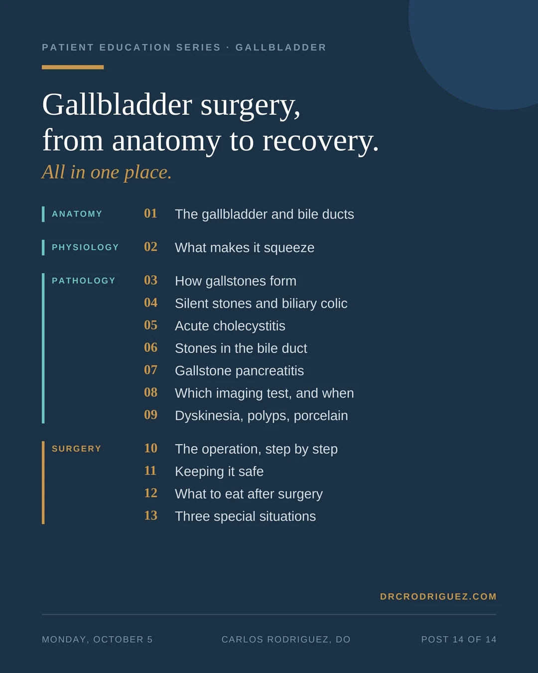
This is the last post in that series. Figure 1 shows all 13 posts on one card. Each one is listed below with a short note on what it covers, so you can start with the question you have today.
Anatomy: what it looks like
Post 1. The gallbladder and bile ducts → Where the liver, gallbladder and bile ducts sit, and how they connect to the intestine. Every post after it builds on this one.
Physiology: how it works
Post 2. What makes the gallbladder squeeze → The liver makes bile and the gallbladder stores it. After a meal, a hormone called cholecystokinin (CCK) tells the gallbladder to squeeze bile into the intestine to help digest fat.
Pathology: what goes wrong
Post 3. How gallstones form → How an imbalance in bile leads to crystals, then sludge, then stones, and who is most at risk.
Post 4. Silent gallstones and biliary colic → Why most gallstones never cause symptoms, and what the pain is like when one does.
Post 5. Acute cholecystitis → What happens when a stone stays stuck at the outlet of the gallbladder, how the inflammation is graded, and why it needs surgery.
Post 6. Stones in the bile duct, and infection → What happens when a stone leaves the gallbladder and blocks the main bile duct, and how an infection behind that blockage is treated.
Post 7. Gallstone pancreatitis → How a stone passing through the bile duct can inflame the pancreas, and how doctors grade how severe it is.
Post 8. Which imaging test, and when → Ultrasound, the HIDA scan (a nuclear medicine test of how well the gallbladder empties), MRCP (an MRI of the bile ducts) and CT, and what each one is good for.
Post 9. Problems that are not stones → Biliary dyskinesia (a gallbladder that does not empty well), polyps (growths on the lining) and porcelain gallbladder (a wall that has turned to calcium).
Surgery: how we fix it
Post 10. The operation, step by step → Four small incisions, the robot, and the order in which I remove the gallbladder.
Post 11. Keeping it safe → The critical view of safety, the dye that makes the bile ducts glow, X-ray pictures of the ducts during surgery, and what I do when inflammation makes the anatomy difficult to discern.
Post 12. What to eat after surgery → Why there is no permanent low-fat diet after this operation, and what we do about diarrhea that does not stop on its own.
Post 13. Three special situations → An inflamed gallbladder with no stones in it, pain that continues after the gallbladder is out, and removing the gallbladder through a single incision.
Bonus posts
Robotic vs. laparoscopic, part 1: the elective case → What the research shows for planned gallbladder surgery.
Robotic vs. laparoscopic, part 2: emergency surgery → What the research shows when the gallbladder comes out urgently.
Part 3: why every gallbladder I take out comes out robotically → Where I land after reading that research.
What I tell patients about recovery → Lifting, showering, driving, and when to call the office.
What comes next
The series also lives on the Guided Series page, where the posts run in order. Next, I start a series on hernias.
References
1. Yokoe M, Hata J, Takada T, et al. Tokyo Guidelines 2018: diagnostic criteria and severity grading of acute cholecystitis (with videos). J Hepatobiliary Pancreat Sci. 2018;25(1):41-54. https://pubmed.ncbi.nlm.nih.gov/29032636/
2. Kiriyama S, Kozaka K, Takada T, et al. Tokyo Guidelines 2018: diagnostic criteria and severity grading of acute cholangitis (with videos). J Hepatobiliary Pancreat Sci. 2018;25(1):17-30. https://pubmed.ncbi.nlm.nih.gov/29032610/
3. Banks PA, Bollen TL, Dervenis C, et al. Classification of acute pancreatitis, 2012: revision of the Atlanta classification and definitions by international consensus. Gut. 2013;62(1):102-111. https://pubmed.ncbi.nlm.nih.gov/23100216/
4. Kumar SK, Shehata DG, Cetrulo LN, et al. SAGES guidelines for the use of intraoperative imaging of the common bile duct. Surg Endosc. 2025;39(11):7091-7102. https://pubmed.ncbi.nlm.nih.gov/40935939/
Dr. Rodriguez served as a speaker and educator for Intuitive Surgical through June 2025. He has no current financial relationship with the company.